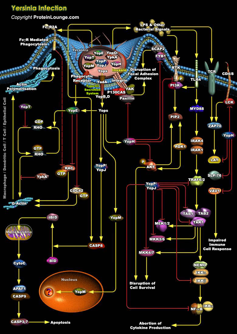
Yersinia pestis, is a facultative intracellular a gram-negative bacillus responsible for causing bubonic plague (Ref.1). Apart from Y. pestis, two other pathogenic Yersinia species, Yersinia enterocolitica, Y. pseudotuberculosis, are known to infect human and animal hosts and cause a variety of intestinal and septicemic diseases. All three species harbor a virulence plasmid, which encodes a type III secretion system (T3SS) for secreting Yop(Yersinia outer membrane proteins) protein substrates, to establish a successful infection (Ref.1). While Y. pestis is responsible for the outbreak of plague, infections with Y. enterocolitica and Y. pseudotuberculosis generally cause gastroenteritis and lymphadenitis (Ref.2). Infection is most often[..]
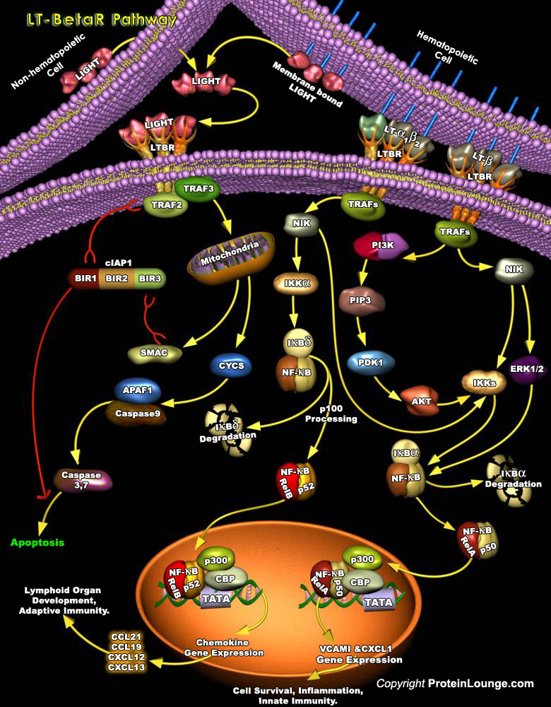
Much of the efficiency of the immune system is attributed to the high degree of spatial and temporal organization in the secondary lymphoid organs. Signaling through the LT-BetaR (Lymphotoxin-Beta Receptor) pathway is a crucial element in the maintenance of this organised microenvironment (Ref.1). LT-BetaR, a member of the TNFR (Tumor Necrosis Factor Receptor) superfamily, plays important roles in embryonic development and organization of secondary lymphoid tissues and maintenance of their architecture in adults (Ref. 2). LT-BetaR is expressed on most cell types including cells of fibroblast, epithelial, and myeloid lineages but not on T or B lymphocytes. It can bind to specific ligands, such as: the membrane form of lymphotoxin heterotrimmer, LT-Alpha1Beta2[..]
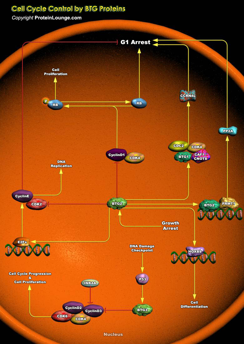
BTG2 (BTG Family Member-2) is endowed with antiproliferative activity. The expression of BTG2 in cycling cells induces accumulation of hypophosphorylated, growth-inhibitory forms of Rb (Retinoblastoma) and led to G1 arrest through impairment of DNA synthesis. Overexpression of CcnD1 (Cyclin-D1) counteracts G1 arrest. Rb is a nuclear phosphoprotein whose phosphorylation state oscillates regularly during the cell cycle. Its under-phosphorylated forms predominate in G0 and G1, while highly phosphorylated forms exist in S, G2 and M phases (Ref.1). The primary biological function of under-phosphorylated Rb is to inhibit progression toward S phase by controlling a checkpoint in late G1. In fact, under-phosphorylated Rb associates with members of the E2F family of[..]
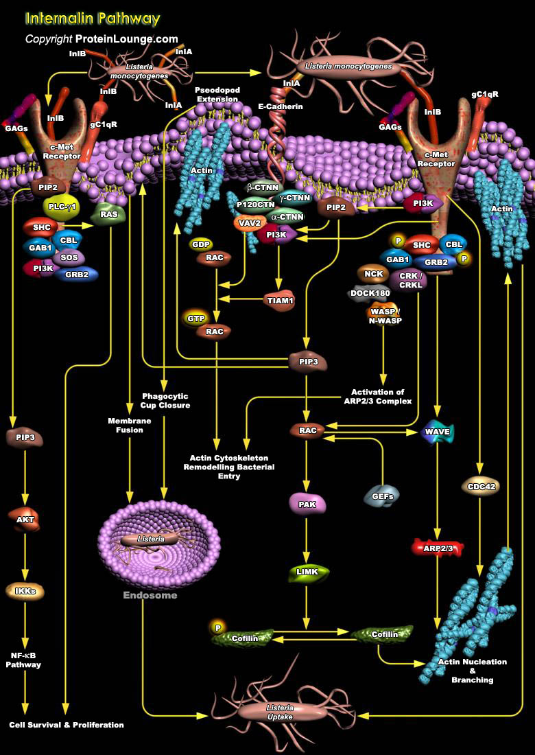
Listeria monocytogenes is a potentially deadly food-borne pathogen that colonizes the gastrointestinal tract of several mammalian species, and can also cause invasive disease and systemic spread if it crosses the intestinal epithelial barrier. Listeria monocytogenes evolved two major molecular invasion proteins, referred to here as invasins: Internalin A (InlA, Internalin) and Internalin B (InlB). These proteins promote internalization into nonphagocytic cells where Listeria monocytogenes can grow in the cytosol as a facultative intracellular pathogen and directly spread to neighboring cells through actin-based motility. InlA-mediated entry is restricted to a few epithelial cells, whereas, InlB promotes entry into various cell types, such as hepatocytes,[..]
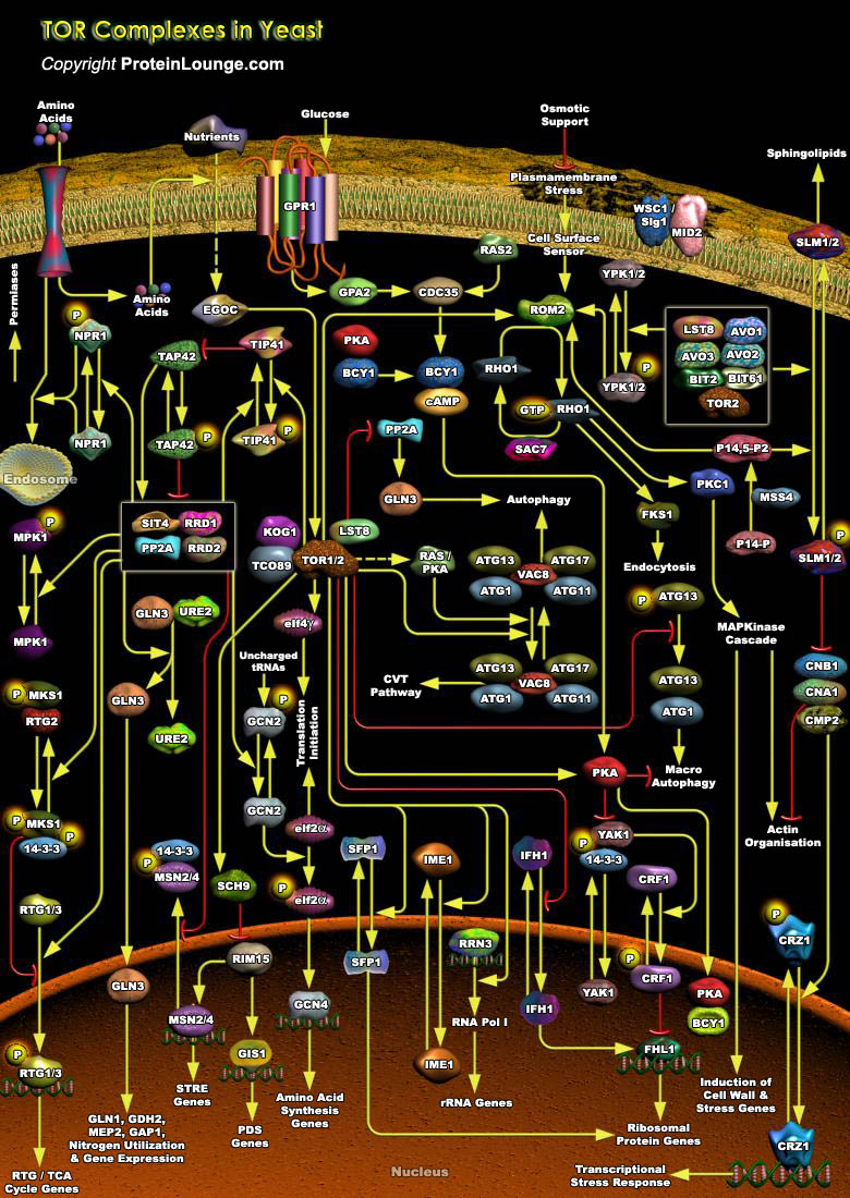
TOR (Target of Rapamycin) is a PIKK (Phosphatidylinositol Kinase-related protein Kinase) that controls cell growth and proliferation. In all eukaryotic cells expressing the protein, TOR function is controlled by nutrient availability, which ensures that protein synthesis is repressed when the supply of precursor amino acids is insufficient. In mammalian cells, one branch of this pathway controls general translational initiation, whereas a separate branch specifically regulates the translation of r-protein (ribosomal protein) mRNAs. In simple organisms, nutrient availability appears to be the major factor influencing TOR activity. In budding yeast, Saccharomyces cerevisiae the TOR pathway similarly regulates general translational initiation, but its specific role in the[..]

Relaxin is a polypeptide hormone that is secreted by the corpus luteum, into the circulation during the menstrual cycle and throughout pregnancy. During the cycle, it stimulates blood vessel growth in the endometrial lining of the uterus during the midluteal phase; coincident with the temporal window during which embryonic implantation occurs. If conception occurs, Relaxin levels rise and remain relatively constant during the entire gestational period (Ref.1). Relaxin is best known for its connective tissue remodeling effects on the female reproductive system. It has diverse actions in the reproductive tract and other tissues during pregnancy. These actions include promotion of growth and dilation of the cervix, growth and quiescence of the uterus, growth and[..]
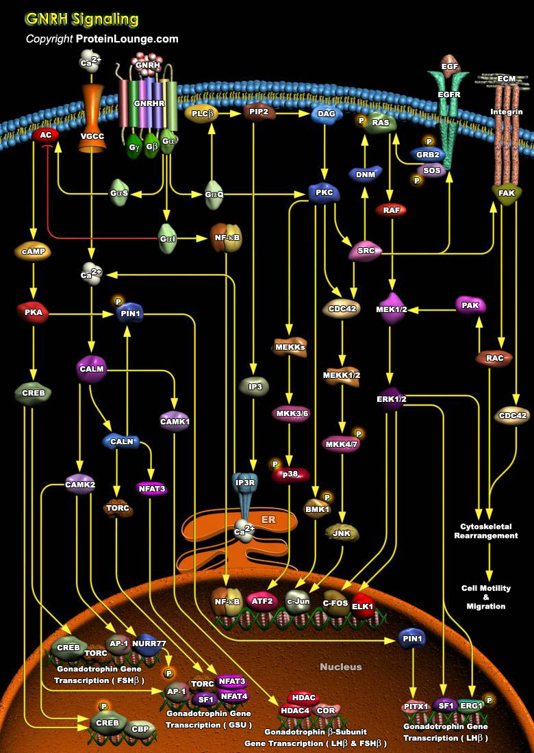
Normal mammalian sexual maturation and reproductive functions require the integration and precise coordination of hormones at the hypothalamic, pituitary, and gonadal levels. The hypothalamic GnRH (Gonadotropin Releasing Hormone), also called LHRH (Luteinizing Hormone Releasing Hormone), is a key regulator in this system (the hypothalamic-pituitary-gonadal axis), that plays a decisive role in the neuroendocrine regulation of human reproduction. GnRH is a decapeptide, released in an episodic manner from the hypothalamic GnRH neurons. The pulsatile delivery of GnRH to the anterior pituitary gland is essential to maintain the circulating gonadotropin profiles. It acts via a specific GPCR (G-Protein Coupled Receptor): GnRHR (GnRH Receptor) and triggers the synthesis of[..]
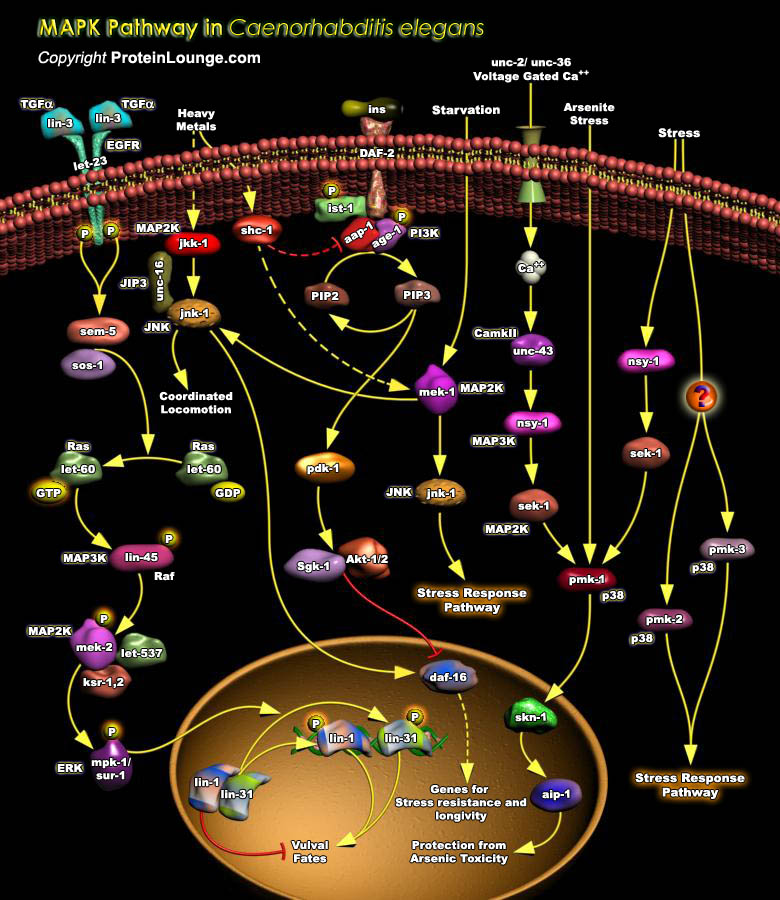
The MAPK (Mitogen-Activated Protein Kinase) pathways are highly conserved signaling cascades that convert extracellular signals into various outputs. Each pathway is composed of three classes of protein kinase: MAPK, MAPKK (MAPK Kinase) and MAPKKK (MAPK Kinase Kinase). MAPK is activated by phosphorylation of specific tyrosine and threonine residues by a family of dual-specificity protein kinase MAPKKs. MAPKK is in turn activated by phosphorylation of serine and serine/threonine residues by a family of upstream MAPKKKs. Each of these upstream components plays a role in multiple cell signaling processes. The ERKs (Extracellular-signal Regulated Kinases), SAPK/JNKs (Stress-Activated Protein Kinases/c-Jun N-terminal Kinases), and p38 are the three best characterized[..]
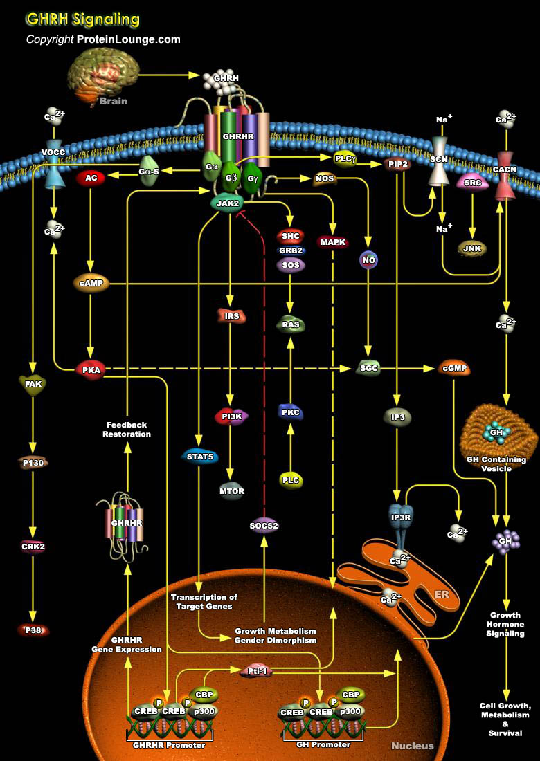
GHRH (Growth Hormone-Releasing Hormone) is a hypothalamic hormone that is essential for normal expansion of the somatotrope lineage during pituitary development. GHRH is produced by GHRH cells in the hypothalamus and reaches the adenohypophysis via the portal system. It stimulates the release of GH (Growth Hormone)/Somatotropin from the adenohypophysis. GH is required for normal postnatal growth, having a critical role in bone growth as well as important regulatory effects on protein, carbohydrate, and lipid metabolism (Ref.1 & 2).GHRH first appears in the human hypothalamus between 18 and 29 weeks of gestation, which corresponds to the appearance of fetal pituitary somatrotropes. It is a 44-amino acid peptide, synthesized by neurons in the hypothalamic arcuate nucleus[..]
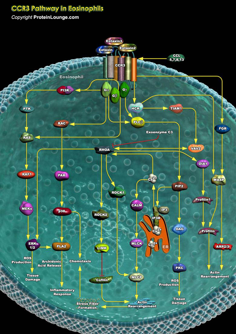
Human eosinophils are crucial effector cells implicated in a number of chronic inflammatory reactions, associated with bronchial asthma, allergic-inflammatory diseases, and parasitic infections.The chemotactic response of eosinophils is mostly mediated by CCR3 (CC Chemokine Receptor-3), a member of the G protein-coupled, seven-transmembrane receptor family, linked to heterotrimeric G-Proteins. Although its expression was first thought to be limited to eosinophils, CCR3 is now known to be more widely expressed on cells involved in allergic inflammation, such as basophils, macrophages, mast cells, neutrophils, airway epithelial cells, and potentially TH2 T-lymphocytes . Chemokines such as: Eotaxin, Eotaxin2, and Eotaxin3 signal exclusively via CCR3 that recruit[..]
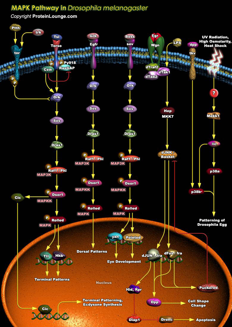
MAPK (Mitogen-Activated Protein Kinase) signal transduction pathways are evolutionarily conserved in eukaryotic cells and transduce signals in response to a variety of extracellular stimuli. Each pathway is composed of three classes of protein kinase: MAPK, MAPKK (MAPK Kinase) and MAPKKK (MAPK Kinase Kinase). MAPK is activated by tyrosine and threonine phosphorylation catalyzed by a family of dual-specificity protein kinase MAPKKs. MAPKK is in turn activated by phosphorylation mediated by MAPKKK. Cascades of MAPKs mediate responses such as cell proliferation, differentiation, and the regulation of metabolic pathways. There are multiple MAPKs in eukaryotes. Three subgroups of the MAPK superfamily have been identified in mammals: ERK (Extracellular signal-Regulated[..]
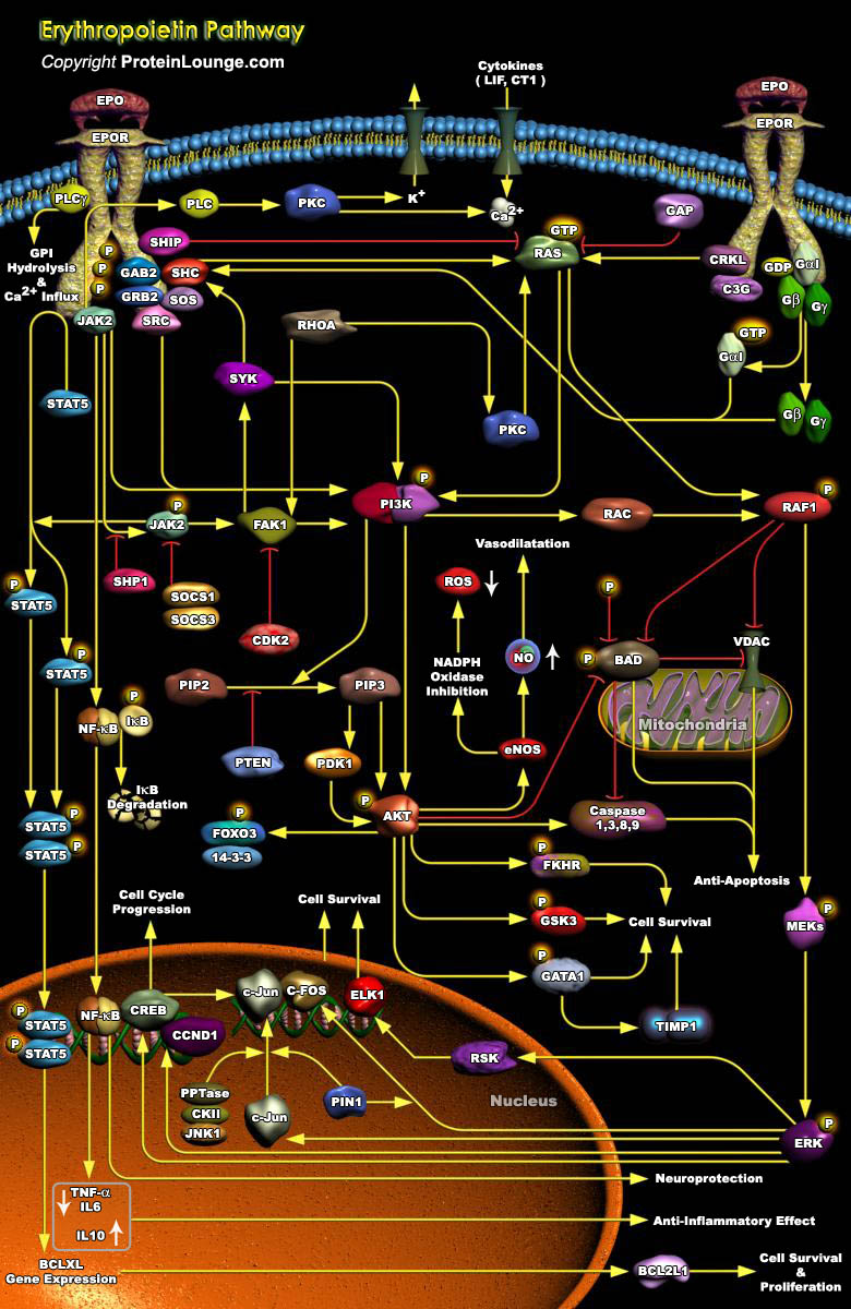
Erythropoiesis is a major pathway for Erythrocyte production, by which pluripotent Hematopoietic Stem Cells give rise to mature end stage cells via a series of differentiations. Epo (Erythropoietin), a glycoprotein hormone and a multifunctional Hematopoietic Cytokine ligand, is the master regulator of Erythropoiesis. As a major function, it monitors the safe passage of the committed Erythroid progenitor cells through several physiological and developmental stages by stimulating Growth, preventing Apoptosis, and promoting terminal differentiation. In addition to its immense survival strategies, Epo initiates Hemoglobin synthesis and is an essential viability and Growth factor in the maintenance of a steady physiological level of circulating Erythrocyte mass which[..]