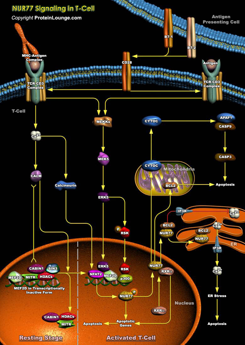
In the immune system, thymus acts as a primary lymphoid organ within which T cells referred to as thymocytes mature. During T cell development in the thymus, immature thymocytes expressing self-reactive TCR (T-Cell Receptor) are eliminated from the developing T-cell repertoire by clonal deletion, or negative selection. This deletion is carried out by Apoptotic signals delivered to thymocytes whose TCR have a high affinity for Self-Antigen/MHC (Major Histocompatibility Complexes)-Antigen Complexes. Negative selection is crucial for generating a peripheral T-lymphocyte population which will recognize only foreign pathogens and helps prevent unwanted immune responses which could lead to autoimmunity (Ref.1). Among the proteins that[..]
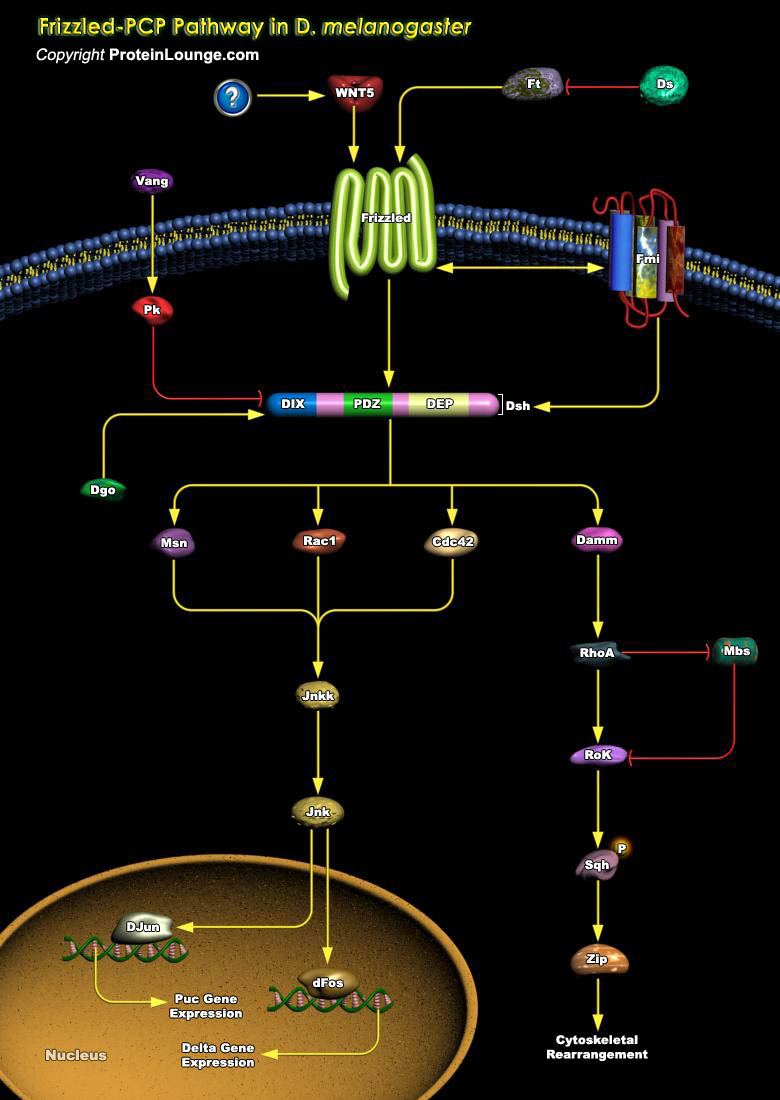
Epithelia are polarized along the apical–basal axis in order to allow for directional transport of proteins within a cell or secretion of factors into. In addition, most epithelia are also polarized within the epithelial plane. The latter polarization is called epithelial PCP (Planar Cell Polarity) or tissue polarity. Planar polarization provides a cell not only with positional information but also directional (vectorial) information and correct PCP is a prerequisite for the formation of many organs. PCP, however, is best studied in Drosophila melanogaster, mainly due to the ease of the fly as a model system. In Drosophila, PCP is externally visible in the alignment of sensory bristles and hairs (trichomes) on the thorax and abdomen, as well as the wing, where[..]
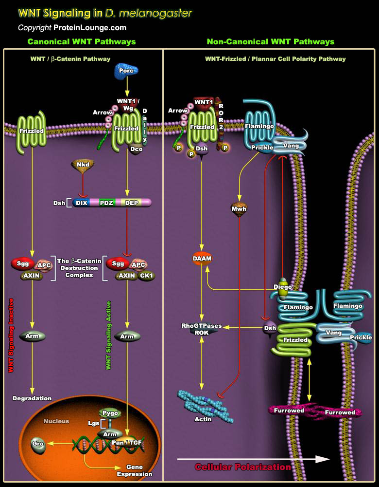
The Wg (wingless) pathway is highly conserved amongst various species and is important for embryonic development, including body axis patterning, cell fate specification, cell proliferation and cell migration. Additionally, Wnt signaling also controls tissue regeneration in adult bone marrow, skin and intestine.Much of our understanding of the role of Wnt proteins during development has come as a result of genetic analyses of the Drosophila Wg gene. In Drosophila, Wg is required during embryogenesis for segmental patterning, muscle development, and midgut formation and to specify appendage primordia. The WNT genes encode a large family of secreted protein growth factors that have been identified in animals from hydra to human. The name WNT denotes the[..]
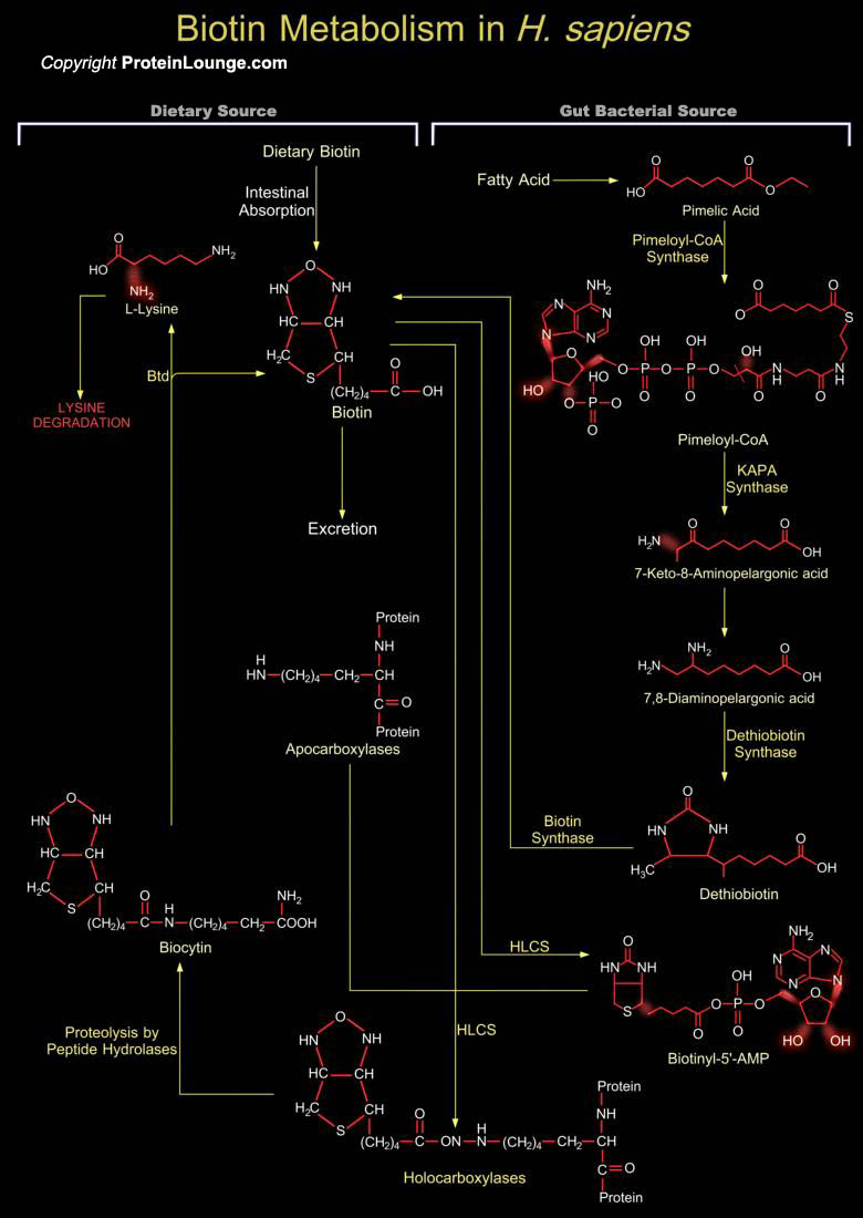
Biotin or Vitamin-H, an essential micronutrient for all mammals belongs to the B-Complex group of Vitamins. It is a water-soluble Vitamin used as co-factor of Biotin-dependent Carboxylases. The role of Biotin in Carboxylases is to act as vector for carboxyl-group transfer between donor and acceptor molecules during Carboxylation reaction (Ref.1). In H. sapiens (Homo sapiens), Biotin is a covalently bound as a prosthetic group for five Biotin-dependent Carboxylases namely PCC (Propionyl-CoA Carboxylase) group; PC (Pyruvate Carboxylase, Mitochondrial); MCC (Methylcrotonyl-Coenzyme-A Carboxylase) group; ACAC-Alpha (Acetyl-Coenzyme-A Carboxylase-Alpha) and ACAC-Beta (Acetyl-Coenzyme-A Carboxylase-Beta). Biotin is covalently attached to Carboxylases by the enzyme HLCS[..]
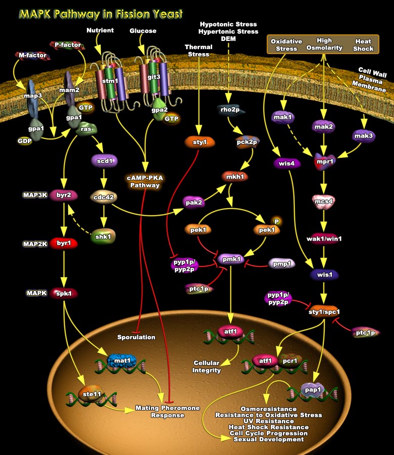
In both Prokaryotes and Eukaryotes, a major adaptive response to various Stress conditions is to change the repertoire of Gene expression. Prokaryotic cells commonly employ the two-component Signal Transduction Systems, where a “sensor” Histidine Kinase, often located in the Plasmamembrane, mediates environmental signals to a cytoplasmic “response regulator” that controls transcription of the target gene. Although Homologous mechanisms have been found also in some Eukaryotic organisms, recent studies have uncovered a pivotal role of MAPK (Mitogen-Activated Protein Kinase) Cascades in stress signaling of Yeast and Vertebrate cells. Each MAPK Cascade comprises a series of three or more protein kinases, each phosphorylating and thereby activating[..]
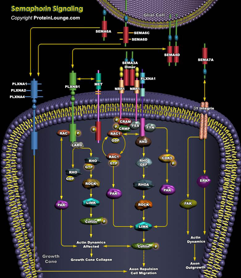
The semaphorins are a family of growth cone guidance molecules, conserved from insects to mammals, which includes proteins strongly implicated in mediating repulsive guidance during neurodevelopment and neuronal regeneration. The range of neurons responsive to semaphorins is extensive and includes dorsal root ganglion, sympathetic, motor, cerebellar, hippocampal, olfactory and corticospinal neurons. Currently, more than 20 members have been identified and they all share a conserved, 500 amino acid residue Sema domain near their amino terminus. Semaphorins are divided into eight classes according to species of origin and structural similarities. The best characterized class of the Semaphorin family is Class 3 Semaphorins. Currently, six members have been identified[..]

Netrin1 (NTN1) Netrin1 is an evolutionarily conserved signaling protein , which functions as a common cue for axonal guidance and vascular patterning and is responsible for integrating regulatory cues from retinal neurons, glia and immune cells.Netrin1 deficiency may result in shunting of metabolic resources to optimize cell survival in viable retinal cells (Ref.1). Exposure to Netrin1 may lead to chemoattraction or chemoreulsion of an axon. The switching responses between attraction and repulsion are dependent on several factors that include levels of Netrin1 receptor on the surface of the growth cone ;activation or inhibition of signaling pathways by intracellular signals like Ca2+, cGMP, or cAMP;relative affinities of different receptor types for Netrin1 as well[..]
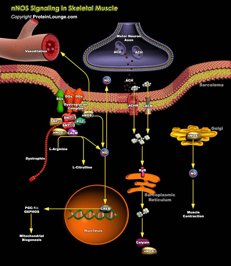
NO (Nitric Oxide) is formed endogenously by a family of enzymes known as NOS (NO Synthases). The distribution of different isoforms of NOS is largely related to their respective functions. Three distinct isoforms of NOS have been identified: nNOS (also known as NOSI and NOS-1) being the isoform first found (and predominating) in neuronal tissue, iNOS (also known as NOSII and NOS-2) which is inducible in a wide range of cells and tissues and eNOS (also known as NOSIII and NOS-3) isoform first found in vascular endothelial cells. NO plays several important roles in the brain, including in regulation of synaptic signaling and plasticity. Additionally, high levels of nNOS protein are present in skeletal muscle, where NO controls muscle contractility and local blood flow[..]
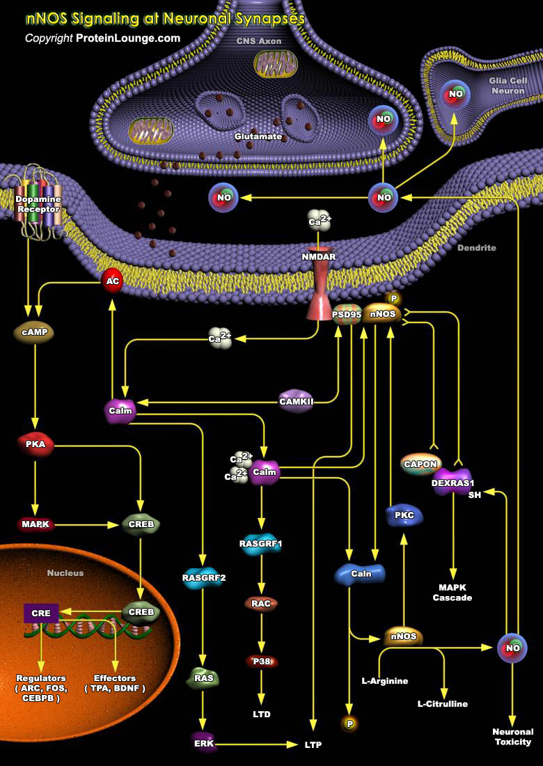
NO (Nitric Oxide) is formed endogenously by a family of enzymes known as NOS (NO Synthases). The distribution of different isoforms of NOS is largely related to their respective functions. Three distinct isoforms of NOS have been identified: nNOS (also known as NOSI and NOS-1) being the isoform first found (and predominating) in neuronal tissue, iNOS (also known as NOSII and NOS-2) which is inducible in a wide range of cells and tissues and eNOS (also known as NOSIII and NOS-3) isoform first found in vascular endothelial cells. Glutamate, the major excitatory neurotransmitter in the brain, is the most effective activator of NO biosynthesis in most brain regions (Ref.1). NO is generated via a five-electron oxidation of a terminal guanidine nitrogen on L-Arginine.[..]
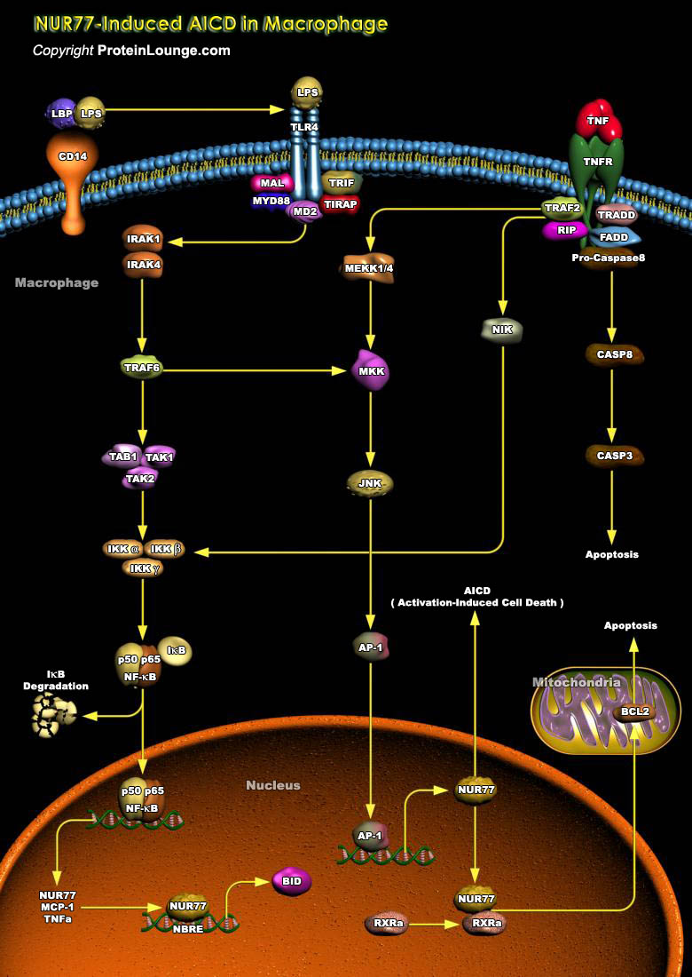
PCD (Programmed Cell Death) or Apoptosis, is a genetically regulated, self-destructive cellular process found in metazoans to eliminate individual cells when they are no longer needed in development, tissue remodeling, or immune regulation, as well as in various diseases. Macrophages are specialized phagocytes responsible for ingesting and digesting senescent, dead, and damaged cells in many tissues as well as microorganisms. These are regarded as very important players in innate immunity because of their nonselective response to almost all infectious microorganisms. Macrophages play such a crucial role in immunity as well as in remodeling tissues, the life span of activated Macrophages is important in physiological and pathological processes. Activated Macrophages[..]
Cell death has been divided into two main types: PCD (Programmed Cell Death), in which the cell plays an active role, and Necrotic (passive) cell death. PCD is a form of cell death in which the cell plays an active role in its own demise. In contrast, Necrosis is characterized by swelling of the cell and its organelles; that results in disruption of the cell membrane and cell lysis. The PCD observed during development and tissue homeostasis has been classified morphologically into three main types: type 1, also known as nuclear or apoptotic; type 2 or Autophagic; and type 3, also referred to as Cytoplasmic. Apoptosis is the best characterized type of PCD, in which the cells display membrane blebbing, flipping of Phosphatidylserine in the plasma membrane, nuclear[..]
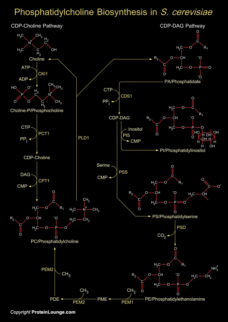
PC (Phosphatidylcholine) is the most abundant Phospholipid in the yeast S. cerevisiae (Saccharomyces cerevisiae) and the major Phospholipid present in eucaryotic cell membranes. It serves as a major structural component of cellular membranes and as a source of several lipid messengers. There are two pathways for PC synthesis in yeast, the CDP-Choline (Cytidine Diphosphate-Choline) pathway (a part of Kennedy Pathway) and the CDP-DAG (Cytidine Diphosphate-Diacylglycerol). Both pathways function to synthesize PC in wild-type cells (Ref.1). The CDP-DAG pathway is used primarily by cells in the absence of exogenous Choline. The PC synthesized by this pathway is constantly metabolized to free Choline and PA (Phosphatidate/Phosphatidic Acid) via the reaction catalyzed by PLD1[..]