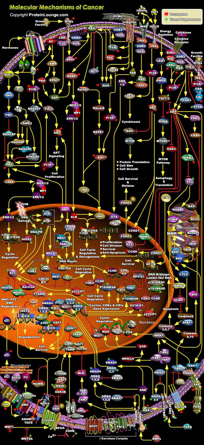
Cancer cell genotypes are a manifestation of six essential alterations in cell physiology that collectively dictate malignant growth; self-sufficiency in growth signals, insensitivity to growth-inhibitory (anti-growth) signals, evasion of programmed cell death (apoptosis), limitless replicative potential, sustained angiogenesis and tissue invasion and metastasis. Environmental and endogenous DNA-damaging agents and genetic instability drive tumor progression by generating mutations in two types of genes, oncogenes and tumor suppressor genes, providing cancer cells with selective growth advantage and thereby leading to the clonal outgrowth of a tumor. In general, oncogenes (called proto-oncogenes in their normal, non-mutated form) promote cell proliferation and[..]
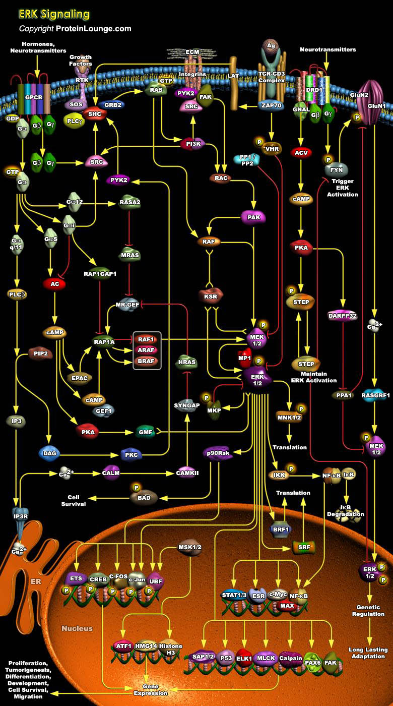
The MAPK (Mitogen-Activated Protein Kinase) pathway is one of the primordial signaling systems that nature has used in several permutations to accomplish an amazing variety of tasks. It exists in all eukaryotes, and controls such fundamental cellular processes as Proliferation, Differentiation, Survival and Apoptosis. Mammalian MAPK can be divided into four groups based on their structure and function: ERKs (Extracellular signal-Regulated Kinases), p38MAPKs, JNKs (c-Jun NH2-terminal Kinases) and ERK5 (Extracellular signal-Regulated Kinase-5) or BMK. Activation of these MAPKs occurs through a cascade of upstream kinases; a MAPKKK (MAPK Kinase Kinase) first phosphorylates a dual-specificity protein kinase MAPKK (MAPK Kinase), which in turn phosphorylates the MAPK. This[..]
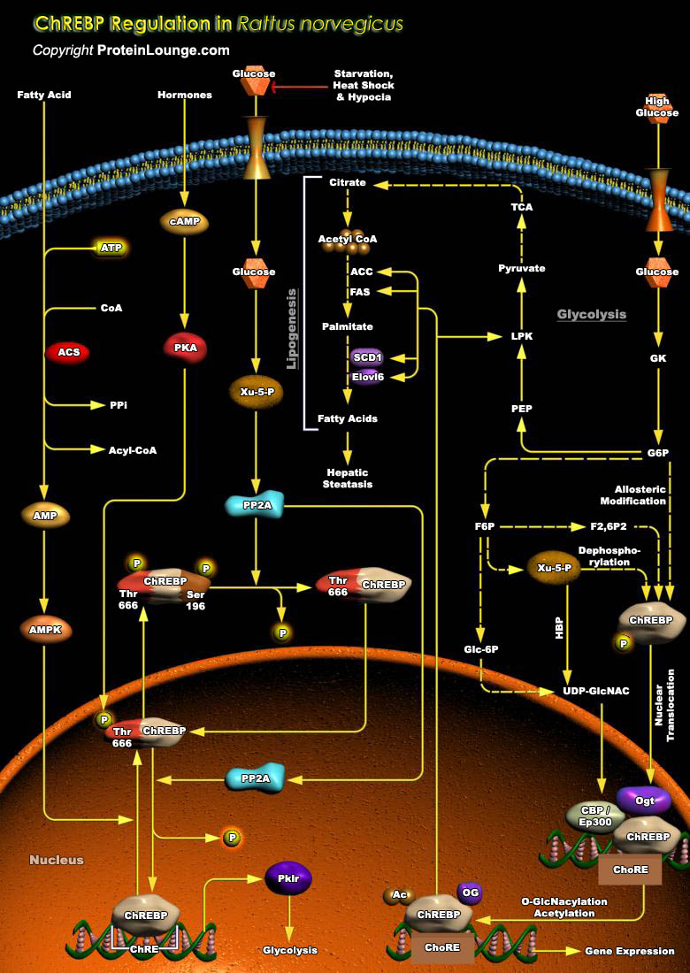
The liver is the major organ responsible for the conversion of excess dietary carbohydrate into triglycerides. Ins (Insulin) and Glucagon (a pancreatic hormone) play critical roles in homeostatsis of Glucose and triglycerides in humans as well as in R. norvegicus (Rattus norvegicus). Glucose serves as a signal independent of hormones in activation of more than 15 enzyme genes in the lipogenic pathways. However, the mechanism by which Glucose generates signal to induce these gene is not known (Ref.1). Ingestion of a high carbohydrate diet leads to the activation of several regulatory enzymes of Glycolysis and Lipogenesis, including the PKLR (Pyruvate Kinase, Liver and RBC), also known as LPK (L-Type Pyruvate Kinase). The lipogenic genes contain ChREs (Carbohydrate[..]
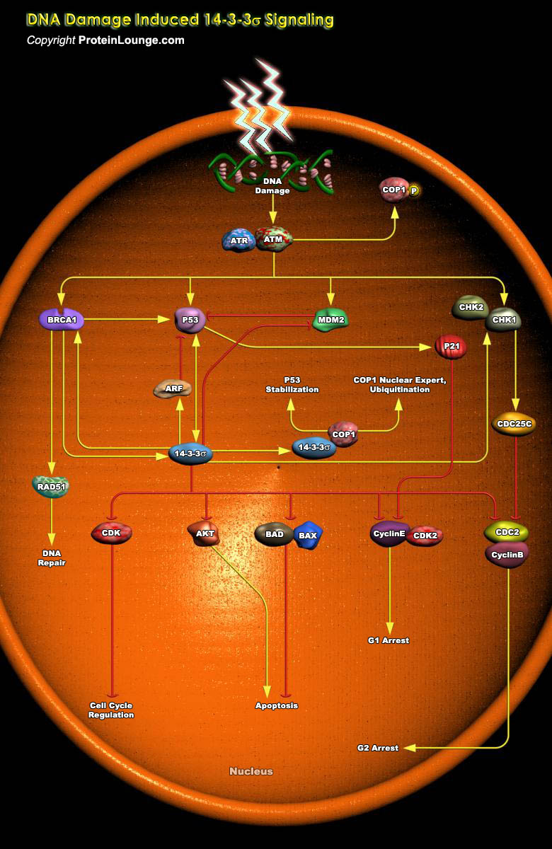
DNA damage checkpoints are critical for preventing tumorigenesis and regulating the response of cells to genotoxic agents. When cells are exposed to DNA damaging agents, they respond by undergoing cell cycle arrest or programmed cell death. 14-3-3 proteins play particularly important roles in coordinating progression of cells through the cell cycle, regulating their response to DNA damage, and influencing life-death decisions following internal injury or external cytokine-mediated cues (Ref.1 and 2). 14-3-3 sigma (also called stratifin or SFN), a tumor suppressor, is a member of a highly conserved family of 14-3-3 proteins that are present in all eukaryotic organisms. Its increased expression causes resistance to anticancer agents and radiation that causes DNA damages[..]
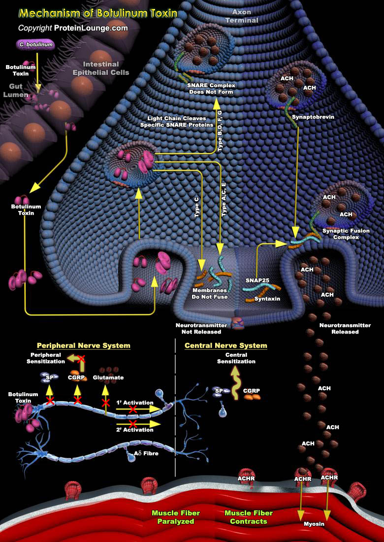
BoNT(Botulinum toxin) is a potent neurotoxin that is produced by the gram-positive, spore-forming, anaerobic bacterium, Clostridum botulinum. There are 7 known immunologically distinct serotypes of BoNT: types A, B, C1, D, E, F, and G. Clostridum neurotoxins are produced as a single inactive polypeptide chain of 150 kDa, which is cleaved by tissue proteinases into an active di-chain molecule: a heavy chain (H) of ~100 kDa and a light chain (L) of ~50 kDa held together by a single disulfide bond. Each serotype demonstrates its own varied mechanisms of action and duration of effect. The heavy chain of each BoNT serotype binds to its specific neuronal ecto-acceptor, whereby, membrane translocation and endocytosis by intracellular synaptic vesicles occurs. The light[..]
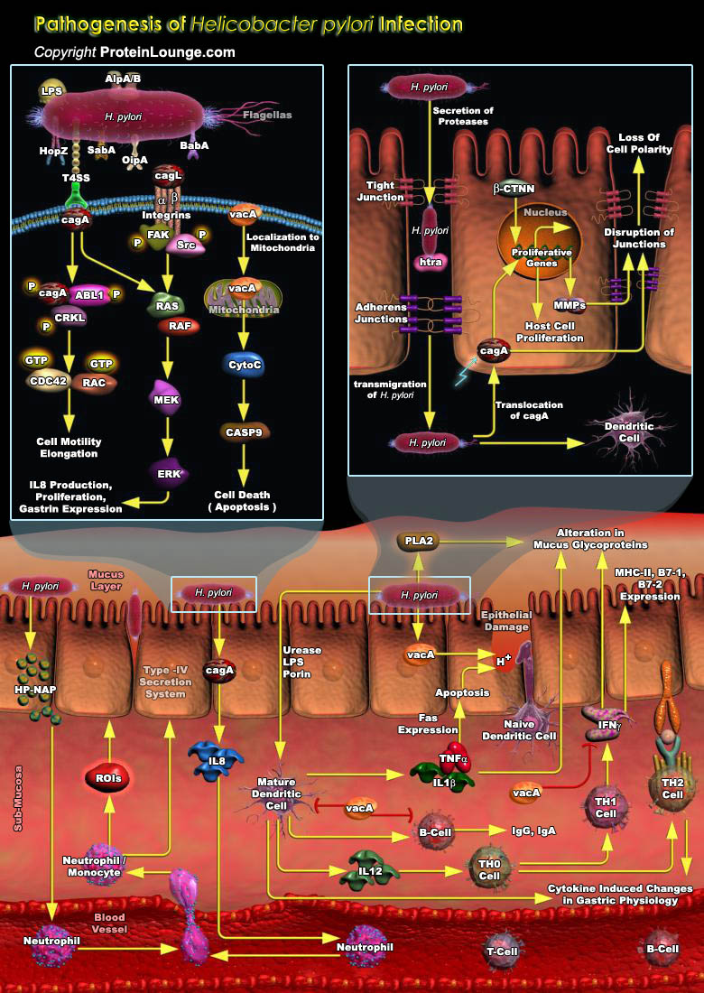
The gastrointestinal tract represents an important barrier between human hosts and microbial populations. One potential consequence of host-microbial interactions is the development of mucosal inflammation. A paradigm for such chronic host-microbial relationships is carriage of Helicobacter pylori, Gram-negative bacteria that colonize the stomachs of humans and primates. H. pylori colonization induces chronic gastritis in essentially all hosts, a process that increases the risk of developing peptic ulceration, distal gastric adenocarcinoma, and gastric mucosal lympho-proliferative disease. However, only a small percentage of persons carrying H. pylori develop clinical sequelae; enhanced risk may be related to differences in expression of specific bacterial products, to[..]

Multiple sclerosis (MS) is an inflammatory, demyelinating, and neurodegenerative disease of the central nervous system that affects both adults and children. MS is characterized by the formation of multiple lesions along the nerve fibers in the brain, spinal cord and optic nerves. Inflammation of the white and gray matter tissues in the CNS due to focal immune cell infiltration and their cytokines are the incipient cause of damage in MS. Hallmark to MS is the demyelinated plaque, which consists of a well-demarcated hypocellular area characterized by the loss of myelin, the formation of astrocytic scars, and the mononuclear cell infiltrates concentrated in perivascular spaces composed of T cells, B lymphocytes, plasma cells, and macrophages. The mechanisms underlying[..]

cAMP (Cyclic Adenosine 3',5'-monophosphate) is the first identified second messenger, which has a fundamental role in the cellular response to many extracellular stimuli. The cAMP signaling pathway controls a diverse range of cellular processes. Indeed, not only did cAMP provide the paradigm for the second messenger concept, but also provided the paradigm for signaling compartmentalization. The different receptors, chiefly the GPCRs (G-Protein Coupled Receptors), Alpha and Beta-ADRs (Adrenergic Receptors), Growth Factor receptors, CRHR (Corticotropin Releasing Hormone Receptor), GcgR (Glucagon Receptor), DCC (Deleted in Colorectal Carcinoma), etc are responsible for cAMP accumulation in cells that cause different physiological outcomes, and changes in cAMP[..]
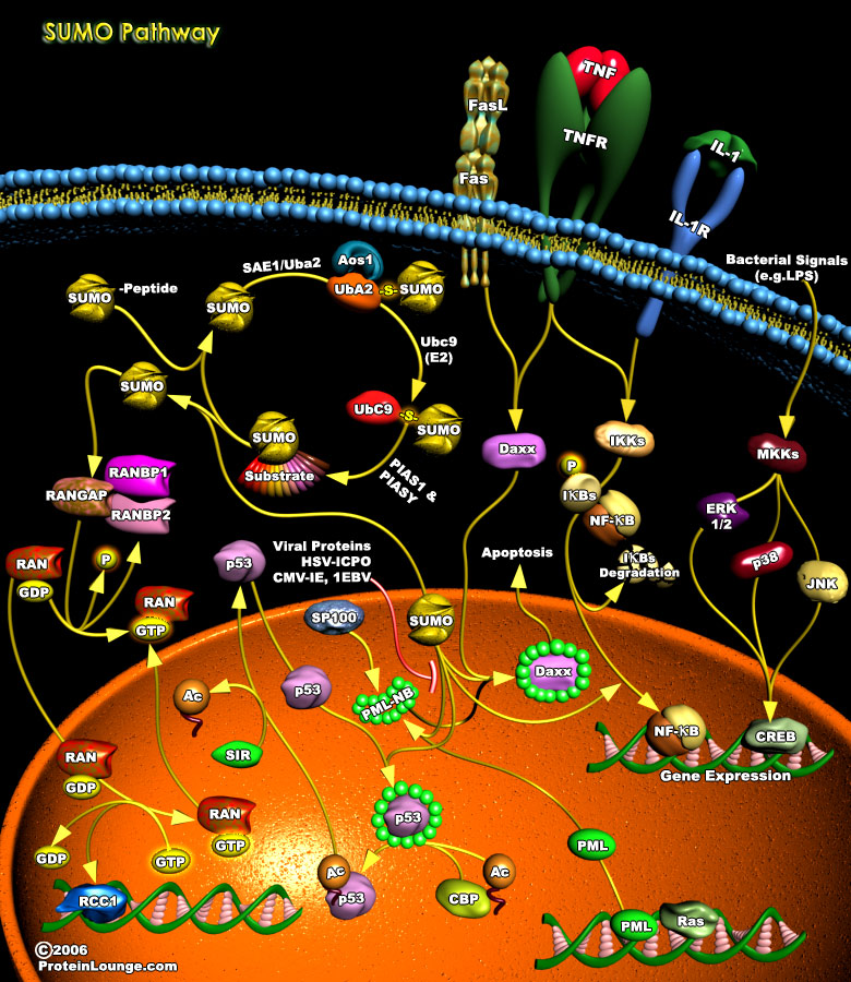
Covalent modifications of proteins, such as phosphorylation, acetylation and ubiquitylation, play an important role in most cellular processes because they can cause rapid changes in the activities of pre-existing proteins. This type of mechanism for regulating protein function is especially crucial in signal transduction pathways and in cell cycle. Posttranslational modifications (PTMs) by members of the ubiquitin family are covalent events that promote radical changes in the properties of modified proteins. Among all ubiquitin-like molecules, a particular attention has been given to the modification by SUMO (Small Ubiquitin Modifier) also known as Sentrin. SUMOylation plays critical roles in a variety of cellular processes, including transcription, cellular[..]
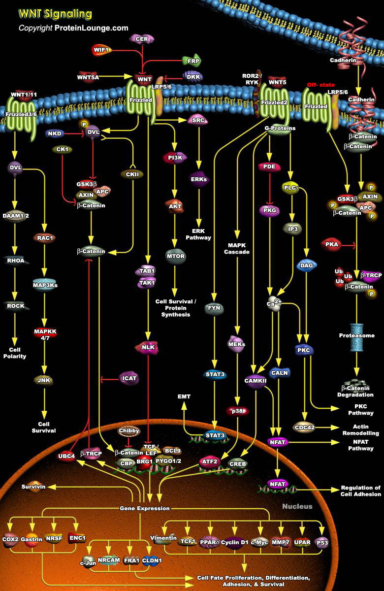
WNT signaling pathways play essential roles in cellular proliferation, differentiation and cell migration during embryonic development. The importance of WNT signaling is indicated by conservation of its molecular components across organisms ranging from nematodes to humans. WNT pathways are classified into canonical WNT/CTNNB or non-canonical (β-catenin-independent) pathways. Canonical WNT/β- catenin signaling is the most studied, and is mediated by nuclear translocation of its central effector CTNNB and Non-canonical WNT signaling occurs independently of CTNNB–TCF/LEF and is stimulated by WNT ligands that bind to a receptor complex of FZD, ROR1/2 or RYK.WNT pathway is implicated in a variety of cancers (Ref.1 and 2).WNT ligands signal via seven[..]
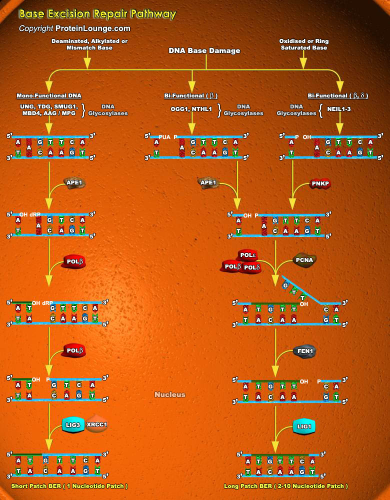
Cells are constantly under threat from the cytotoxic and mutagenic effects of DNA damaging agents that result from either endogenous sources (cellular metabolic processes) or exogenous sources (environmental factors). Endogenous sources of DNA damage include hydrolysis, oxidation, alkylation, and mismatch of DNA bases; sources for exogenous DNA damage include ionizing radiation (IR), ultraviolet (UV) radiation, and various chemicals agents (Ref.1). Repairing damage in DNA from anything that causes a mutation, such as UV radiation and tobacco smoke, is a fundamental process that protects our cells from becoming cancerous. The major forms of DNA damage include SSB (Single-strand Breaks), DSB (Double-strand Breaks), alteration of bases, hydrolytic depurination, hydrolytic[..]
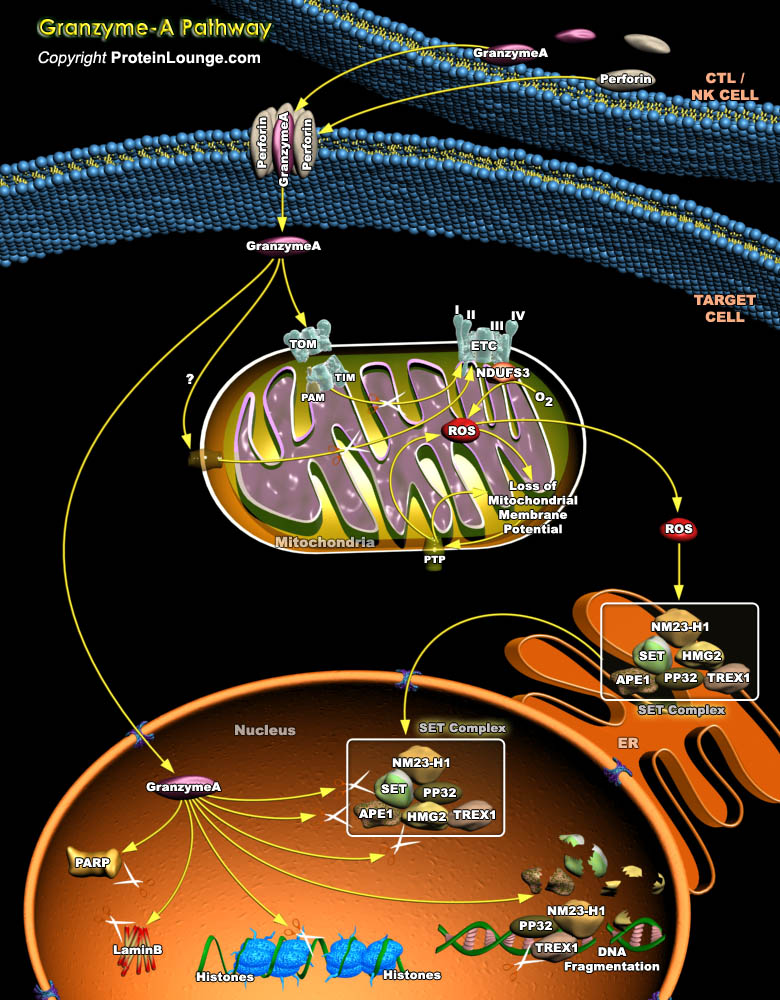
CTLs (Cytotoxic T Lymphocytes) and NK (Natural Killer) cells are the key immune effectors that eradicate infected cells or tumors. To destroy these targets, CTLs and NK cells mostly use the granule exocytosis pathway, which releases perforin and Granzymes from cytolytic granules into the immunological synapse formed with the target. Granzyme-A and Granzyme-B, the most abundant Granzymes, are delivered to the target cell cytosol through perforin and independently induce cell death. The tryptase Granzyme-A activates cell death through a caspase-independent mechanism. Granzyme-A causes characteristic features of apoptosis, including membrane blebbing, loss of mitochondrial transmembrane potential, nuclear fragmentation and chromatin condensation; however, instead of[..]