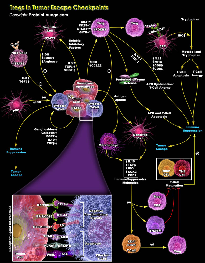
Specific T cell populations have suppressive/regulatory cells known as Regulatory T-cell or Tregs (Previously known as suppressor T-cell). Among them CD4+ regulatory T cells (Tregs) basically has two different subsets Tr1 and Th3 cells which are differentiated by their distinct suppressive mechanisms. The thymus-derived Tregs or natural Tregs (nTregs) express CD4 and high CD25 with FOXP3 (Forkhead Box-P3) to be a key regulatory gene. Apart from these, other markers of Treg include CTLA4 (Cytotoxic T-Lymphocyte-Associated protein-4), GITR (Glucocorticoid-Induced TNFR-Related protein) and LAG-3 (Lymphocyte Activation Gene-3). Treg cells are implicated in the development of autoimmunity, allergy and rejection of organ transplants, as well as the suppression of immune[..]
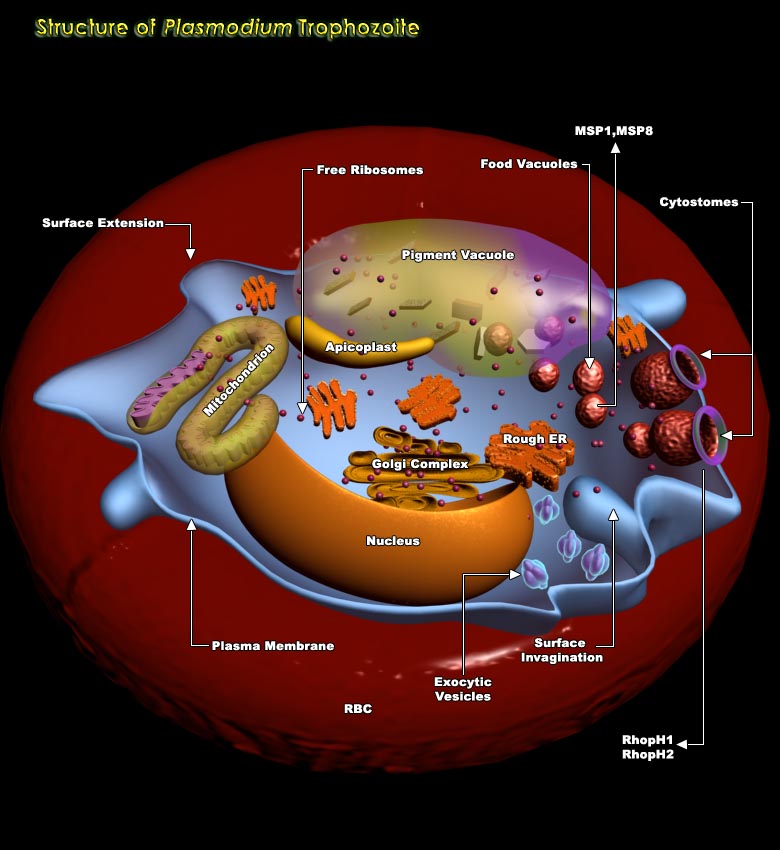
In the life cycle of Plasmodium spp, Erythrocytic stage is very important involving four stages viz. Merozoite stage, ring stage, Trophozoite stage and Schizont stage. On being released from the hepatocytes, the Merozoites enter the bloodstream prior to infecting red blood cells. They use the Apicomplexan invasion organelles to recognize and enter the host erythrocyte. The parasite first binds to the erythrocyte in a random orientation. It then reorients such that the apical complex is in proximity to the erythrocyte membrane. A tight junction is formed between the parasite and erythrocyte. As it enters the red blood cell, the parasite forms a parasitophorous vesicle, to allow for its development inside the erythrocyte. After invading the erythrocyte, the parasite[..]
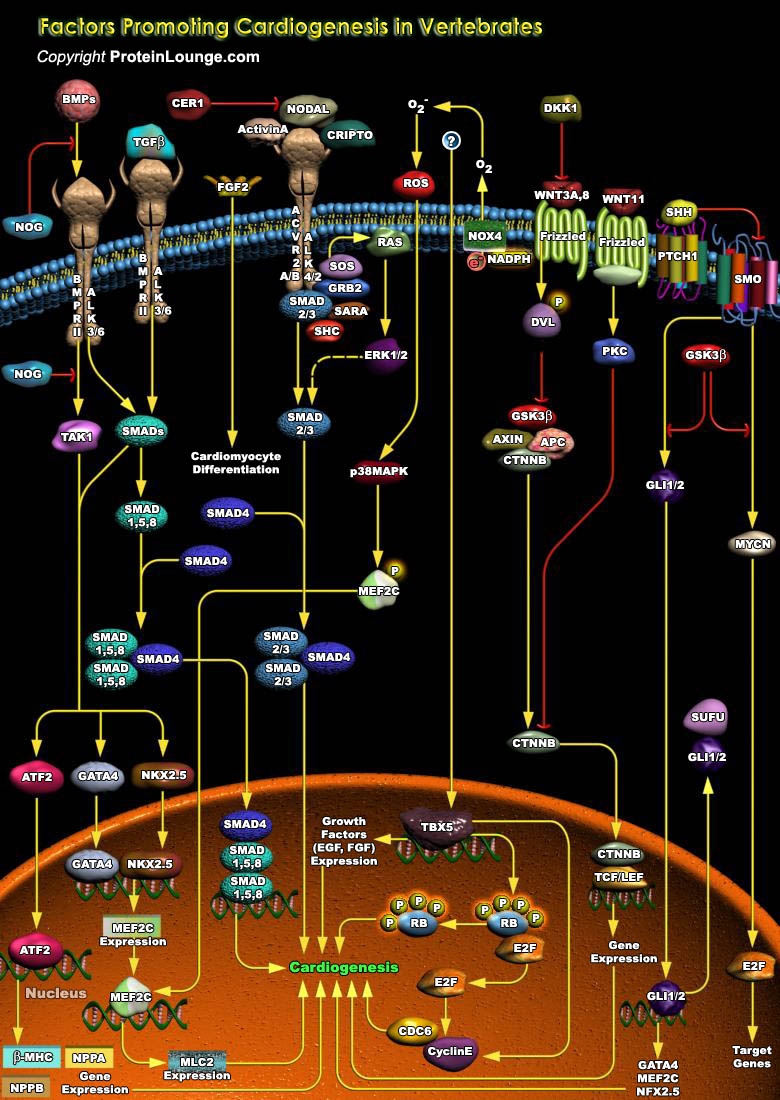
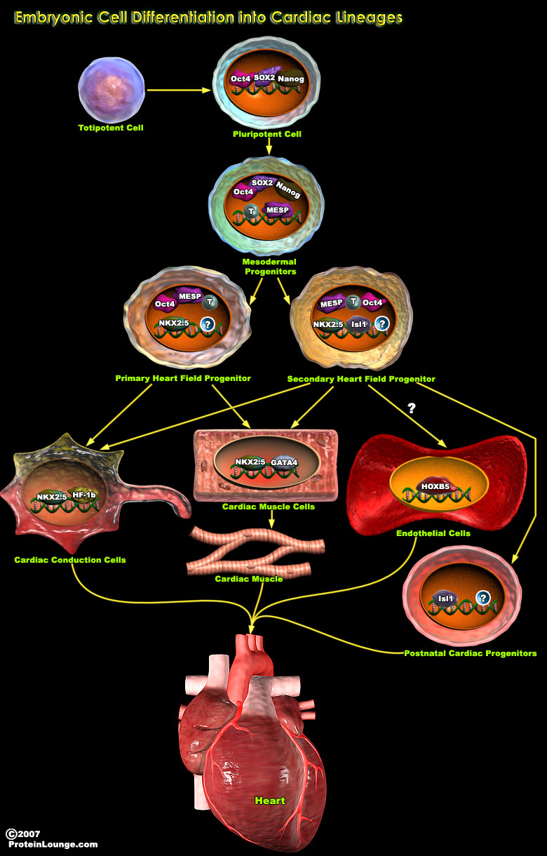
Cardiogenesis is the formation of new heart tissue from embryonic, postnatal, or adult multipotent cardiovascular progenitor cells. Cardiogenesis in vertebrate is a complex process, where various genetic and epigenetic factors play crucial role in driving the interaction between different structures and diverse cell types. Cardiomyocytes are the main cell type found in the heart that are responsible for the contraction of the chambers and efficient blood flow throughout the body (Ref.1 and 2). Multiple transcription factors and extracellular growth factors specify the Cardiac lineage in mesodermal progenitor cells. The earliest expressed transcription factors that initiate Cardiac fate are the homeobox transcription factor NKX2.5 (NK2 Transcription Factor Related[..]
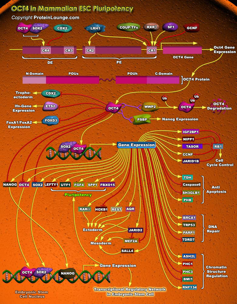
Stem cells are undifferentiated cells capable of producing virtually all cell types in our body. They are characterized by the ability to Self-renew and maintain Pluripotency. For proper developmental outcome, ESCs (Embryonic Stem Cells) must tightly regulate their differentiation status. Hundreds of genes have been identified, including several transcription factors, which have expression patterns tightly correlated with ESC differentiation. Very low number of transcription factors is responsible for regulating the development of an entire organism. These transcription factors form multiprotein complexes on DNA, thereby orchestrating the correct temporal–spatial expression of developmental genes. The process leads to the establishment of functional[..]
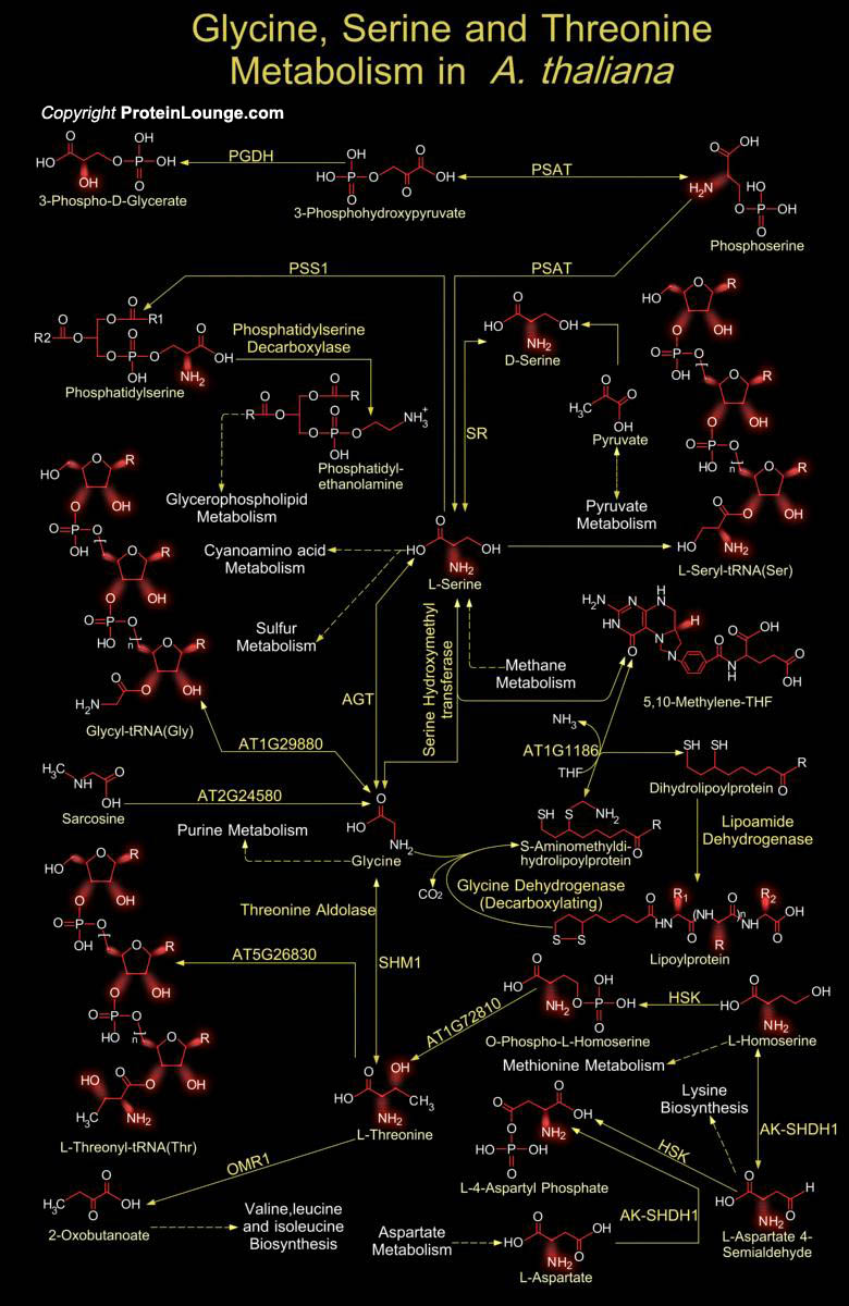
Amino acids are not only the building blocks of proteins, but also serve as precursors for other important plant metabolites and constitute an essential part of human and animal diets. The biosynthesis and degradation of the twenty standard Amino acids represent the complexity and ingenuity of metabolism at its most astounding. Plants are able to generate all 20 Amino acids necessary for protein synthesis by themselves. They do even synthesize some more Amino acids. Glycine and Serine are two Interconvertible Amino acids that play an important role in C1 metabolism. Glycine is the simplest Amino acid and is the only Amino acid that is not optically active. On a molar basis, Glycine is the second most common Amino acid found in proteins and enzymes being[..]
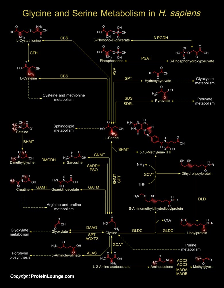
Glycine and Serine are two non-essential amino acids in humans, which have important roles in the Central Nervous System. Serine, a constituent of brain proteins and nerve coverings, is important in various processes like dendritic outgrowth, formation of cell membranes, metabolism of Purines and Pyrimidines, Myelin formation and muscle synthesis, synthesis of nucleotides and neuroactive amino acids like D-Serine and Glycine. As a building block of proteins and membrane lipids, it is required for metabolism of fats, cell and tissue growth and in the immune system, it assists in the production of immunoglobulins/antibodies. Serine derivatives are important components of the membrane phospholipids, whereas Glycine is the simplest, optically inactive, and the second most[..]
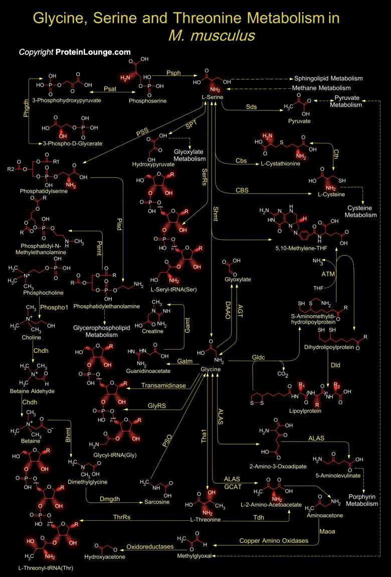
Glycine and Serine are two non-essential neurotrophic amino acids that play an essential role in neuronal development and function in M. musculus (Mus musculus). They share similar neurotrophic effects in promoting neuronal survival and differentiation of sensory ganglia, hippocampal neurons, and cerebellar Purkinje cells. In M. musculus, interplay between Glycine and Serine in a co-existence with L-Threonine represent a major metabolic crossroad that links several other biological pathways of immense importance. L-Serine serves as a building block of proteins, and is of vital importance for the syntheses of L-Cysteine, Phosphatidyl-L-Serine, nucleotides, and the neuromodulators D-Serine and Glycine (Ref.1). L-Serine is a precursor for the synthesis of membrane[..]
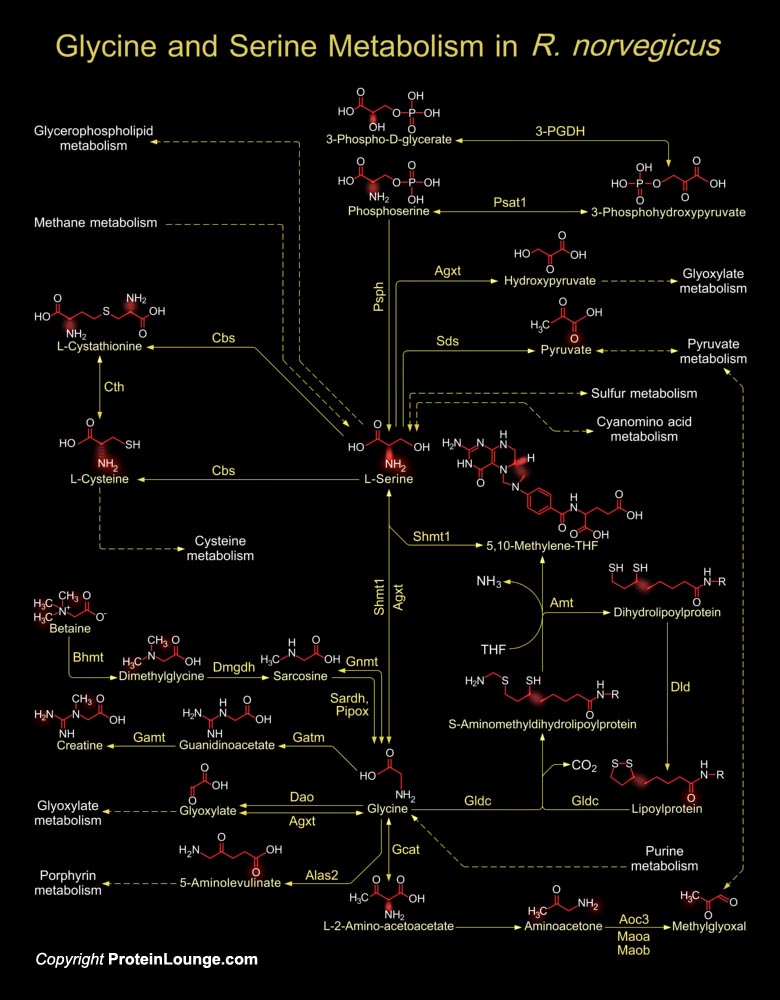
Glycine and Serine are two interconvertible non-essential amino acids found abundantly in almost all cell types. They serve as active ligands for many metabolic pathways where they aid in the synthesis of other essential metabolites, such as Glycogen, Glyoxylate and Pyruvate, which are of immense importance for many cellular and biological processes. The metabolic pathway that correlates Glycine and Serine in R. norvegicus (Rattus norvegicus) has a relation with metabolism of the indispensable amino acid Threonine (Ref.1).L-Serine is a major source of one-carbon units in mammals. It serves as a building block for protein synthesis and is modified in different metabolic pathways for the generation of several essential compounds, such as Glycine, D-Serine, Cysteine,[..]
MI(Myocardial infarction ) causes the loss of cardiac tissue and scar formation, which ultimately lead to heart failure. According to the World Health Organization, heart failure initiated by MI and coronary artery disease accounts for 29% of deaths worldwide. However, human heart tissue does not regenerate spontaneously, thus “regenerative medicine” represents a promising alternative treatment for MI. Cardiac tissue regenerative medicine involves cardiomyocyte regeneration, neovascularization, and paracrine cytokines, which have anti-inflammatory, anti-apoptotic, and anti-remodeling effects. During the last decade, stem cells have become promising candidates for regenerative medicine not only because of their capacity of differentiation toward[..]
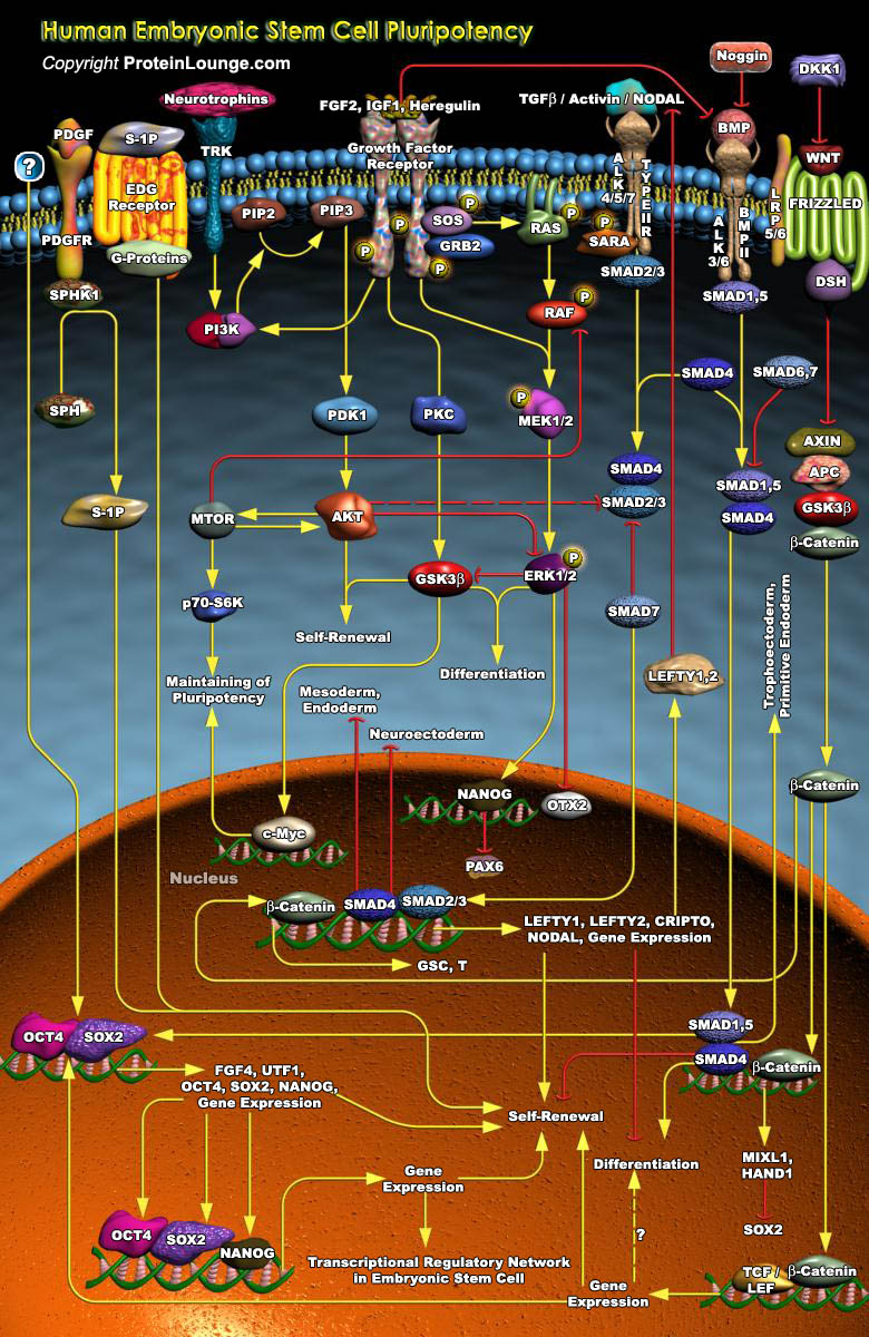
ESCs (Embryonic Stem Cells) are Pluripotent cells capable of differentiating into any cell type of the body. Only three species of Mammals have yielded long-term cultures of self-renewing ESCs- Mice, Monkeys, and Humans. Human ESCs are derived from Blastocysts, multicellular structures originating from four cleavages of fertilized oocytes. Isolated from the ICM (Inner Cell Mass) of Blastocysts, the ESCs retain properties of self-renewal and the potential to be committed and to differentiate toward most cell lineages. They are able to spontaneously give rise to different progenies of the three embryonic layers, namely, the Ectoderm, the Mesoderm and the Endoderm. The Pluripotency of ESCs has attracted great attention for their potential use in tissue and cell therapy.[..]
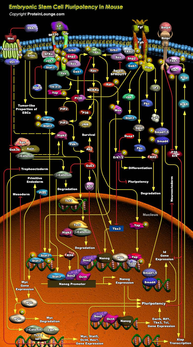
ESCs (Embryonic Stem Cells) are a population of Pluripotent, Self-renewing cells which can proliferate indefinitely and contribute to the formation of basically all cell types in vitro and in vivo. The study of mammalian ESCs, especially Mouse ESCs, has provided valuable insights into early embryogenesis in mammals. Mouse ES cells are derived mainly from the ICM (Inner Cell Mass) of the Mouse Blastocyst embryos and retain this developmental identity even after prolonged culture in vitro. Both cell Extrinsic and Intrinsic factors regulate Mouse ESC Self-renewal and Pluripotency. Cell extrinsic factors include LIF (Leukaemia Inhibitory Factor) and BMP (Bone Morphogenic Protein), which signal through STAT (Signal Transducer and Activator of Transcription) and SMAD (Sma[..]
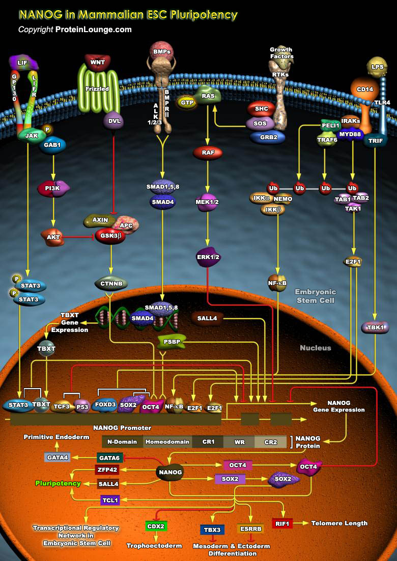
ESCs (Embryonic stem cells) are Pluripotent cells derived from the ICM (Inner Cell Mass) of Blastocyst-stage embryos. These cells have two distinctive properties: an unlimited capacity for Self-renewal and Pluripotency. The capability for Self-renewal and the Pluripotency of ESCs seem to be under the control of multiple transcriptional factors, most common among them being Nanog (Nanog homeobox), Oct4 (Octamer Binding Transcription Factor-4) and SOX2 (SRY (Sex Determining Region-Y) Box-2). Functions of these transcription factors depend on the stage of development of a Pluripotent cell, indicating that these factors function in combination with other processes. The activity of these transcription factors also depends on the accessibility[..]