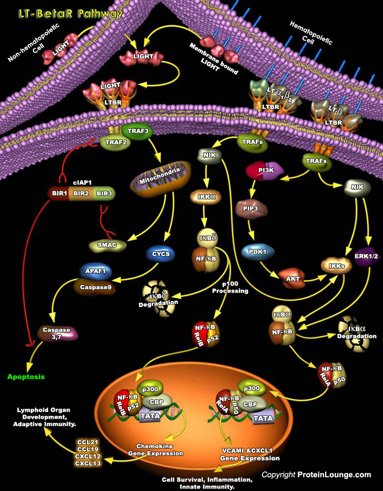
Much of the efficiency of the immune system is attributed to the high degree of spatial and temporal organization in the secondary lymphoid organs. Signaling through the LT-BetaR (Lymphotoxin-Beta Receptor) pathway is a crucial element in the maintenance of this organised microenvironment (Ref.1). LT-BetaR, a member of the TNFR (Tumor Necrosis Factor Receptor) superfamily, plays important roles in embryonic development and organization of secondary lymphoid tissues and maintenance of their architecture in adults (Ref. 2). LT-BetaR is expressed on most cell types including cells of fibroblast, epithelial, and myeloid lineages but not on T or B lymphocytes. It can bind to specific ligands, such as: the membrane form of lymphotoxin heterotrimmer, LT-Alpha1Beta2[..]
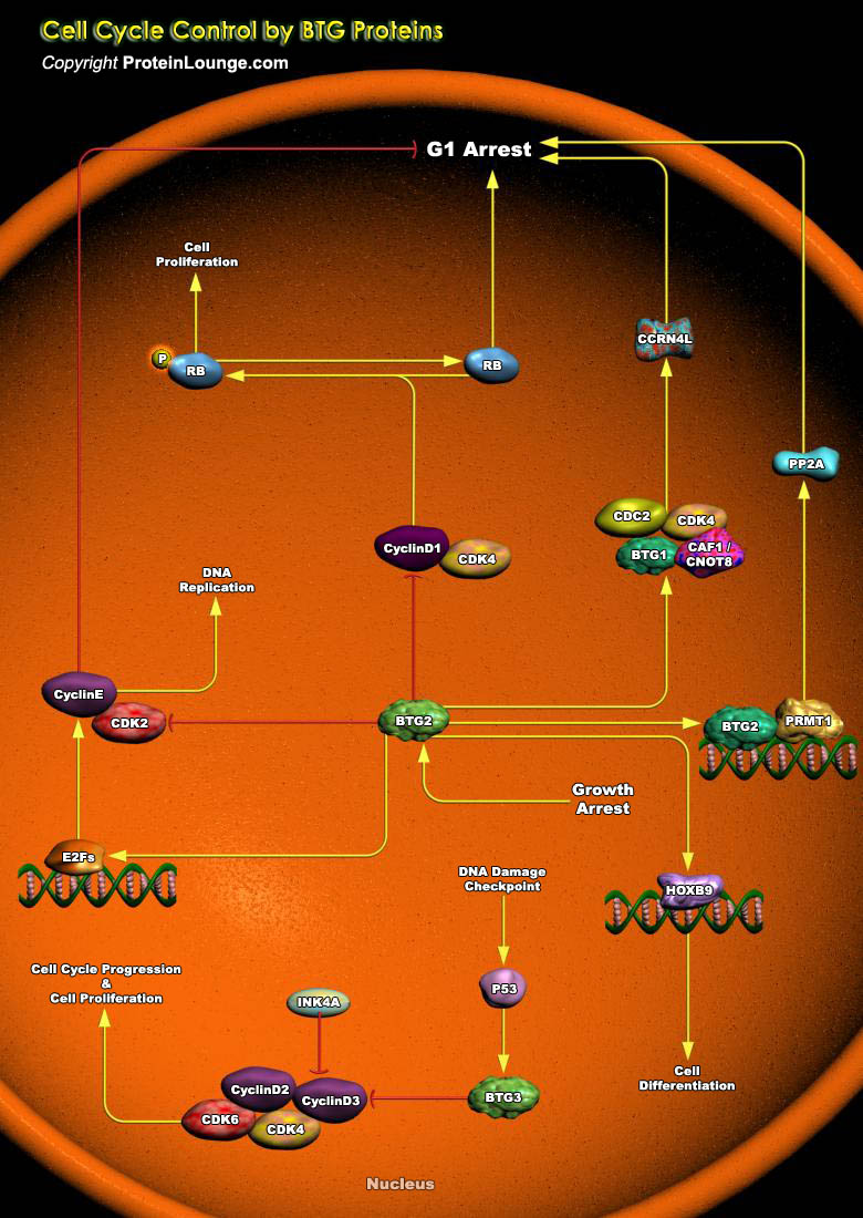
BTG2 (BTG Family Member-2) is endowed with antiproliferative activity. The expression of BTG2 in cycling cells induces accumulation of hypophosphorylated, growth-inhibitory forms of Rb (Retinoblastoma) and led to G1 arrest through impairment of DNA synthesis. Overexpression of CcnD1 (Cyclin-D1) counteracts G1 arrest. Rb is a nuclear phosphoprotein whose phosphorylation state oscillates regularly during the cell cycle. Its under-phosphorylated forms predominate in G0 and G1, while highly phosphorylated forms exist in S, G2 and M phases (Ref.1). The primary biological function of under-phosphorylated Rb is to inhibit progression toward S phase by controlling a checkpoint in late G1. In fact, under-phosphorylated Rb associates with members of the E2F family of[..]
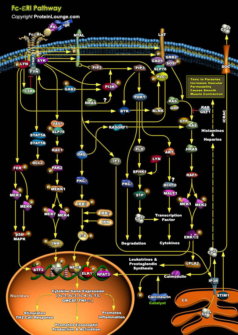
FcεRI is constitutively expressed on mast cells as a tetrameric receptor composed of the IgE-binding α chain, the membrane-tetraspanning β chain, and the disulfide-linked homodimer of the γ chains. The level of expression of the FcεRI on the mast cell surface can be influenced by several factors, such as IgE availability or IgE binding. Antigen (Ag) ligation of IgE-bound FcεRI initiates phosphorylation cascades that cause profound morphological and transcriptional modifications. As FcεRI lacks intrinsic tyrosine kinase activity, its activation requires the downstream phosphorylation of several Src kinases (LYN, SYK, and FYN) (Ref.1).Allergic rhinitis, asthma, atopic dermatitis, and food and drug allergies, which in some cases can lead[..]
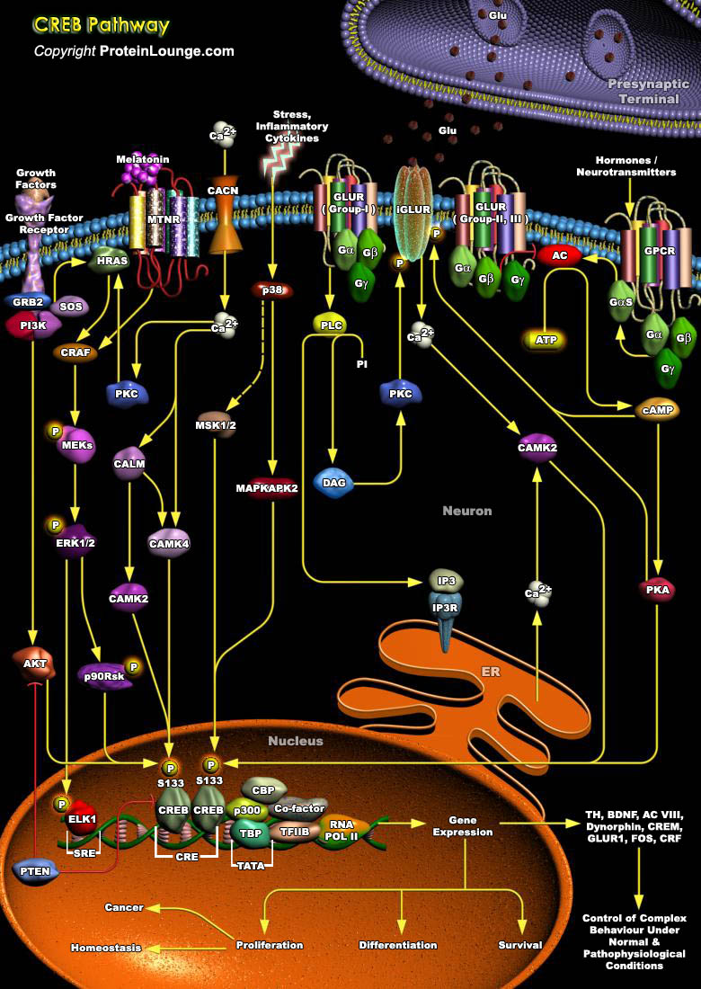
The process of consolidating a new memory and the dynamic complexity of information processing within neuronal networks is greatly increased by activity-dependent changes in gene expression within individual neurons. A leading paradigm of such regulation is the activation of the nuclear transcription factor CREB (cAMP Responsive Element Binding Protein), and its family members the ATF (Activating Transcription Factor) and CREM (cAMP Response Element Modulator), which belong to bZIP (basic/leucine zipper) class of transcription factors that functions in vivo to regulate the proliferation of pituitary cells and thymocytes. Proteins belonging to this class are characterized by the ability to bind to the consensus sequence TGACGTCA (Ref.1, 2 & 3) and contain a leucine[..]
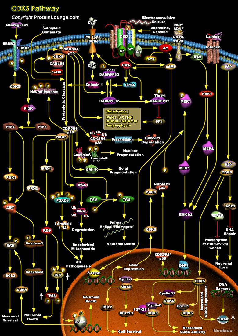
CDKs (Cyclin-dependent kinases) are a group of serine/threonine protein kinases activated by binding to a regulatory subunit cyclin.CDK5(Cyclin dependent kinase-5), a family member of the cyclin-dependent kinases, plays a pivotal role in the central nervous system. During embryogenesis, CDK5 is indispensable for brain development and, in the adult brain, it is essential for numerous neuronal processes, including higher cognitive functions such as learning and memory formation. However, CDK5 activity becomes deregulated in several neurological disorders, such as Alzheimer's disease, Parkinson's disease and Huntington's disease, which leads to neurotoxicity. Therefore, precise control over CDK5 activity is essential for its physiological functions. For its activation[..]
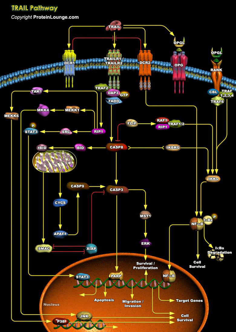
TRAIL (TNF-Related Apoptosis-Inducing Ligand) is a protein consisting of 281 amino acids. It is also called Apo2L. Five proteins, TRAILR1 (DR4), TRAILR2 (DR5/ TRICK2 or KILLER), TRAILR3 (DcR1/ TRID or LIT), TRAILR4 (DcR2 or TRUNDD), and Opg (Osteoprotegerin), have been identified as TRAIL receptors. Both TRAILR1 and TRAILR2 contain the functional DD (Death Domain), capable of inducing apoptosis. The other three receptors DcR1, DcR2 and Opg serve as "decoy" receptors. These three receptors can bind to TRAIL, but cannot induce apoptosis. DcR1 is a glycosylphosphatidylinositol-anchored cell surface protein, which contains the TRAIL-binding region as well as a region that anchors the receptor to the membrane. But unlike the other receptors, DcR1 lacks an[..]
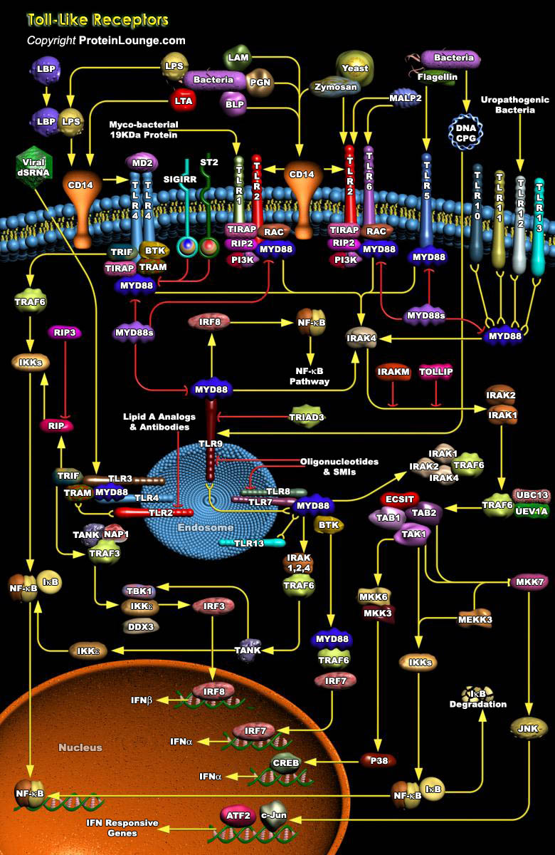
TLRs (Toll-like receptors) are transmembrane proteins expressed by cells of the innate immune system, which recognize invading microbes and activate signaling pathways that launch immune and inflammatory responses to destroy the invaders. Toll receptors were first identified in Drosophila. In mammals, the TLR family includes eleven proteins (TLR1-TLR11). Recently, two new members, TLR12 and TLR13 have been discovered in mouse, but not much information is known about them. Mammalian TLRs consist of an extracellular portion containing Leucine-rich repeats, a transmembrane region and a cytoplasmic tail, called the TIR (Toll-IL-1R (Interleukin-1-Receptor)) homology domain. Different TLRs serve as receptors for diverse ligands including Bacterial cell wall components, viral[..]
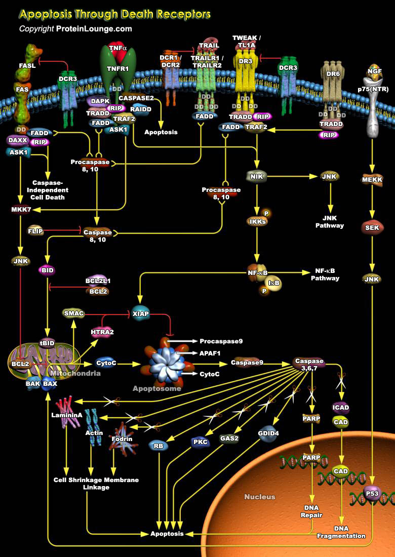
Apoptosis is a cell suicide mechanism that enables metazoans to control cell number in tissues and to eliminate individual cells that threaten the animal's survival. Certain cells have unique sensors, termed DR (Death Receptors), on their surface, which detect the presence of extracellular death signals and, in response; they rapidly ignite the cell's intrinsic apoptosis machinery. Death Receptors belong to the TNF (Tumour Necrosis Factor) gene superfamily and generally can have several functions other than initiating apoptosis. Eight members of the Death Receptor family have been characterized so far: TNFR1 (Tumor Necrosis Factor Receptor-1) also known as DR1, CD120a, p55 and p60, Fas (also known as DR2, APO1 and CD95), DR3 (Death Receptor-3) (also known as APO-3,[..]
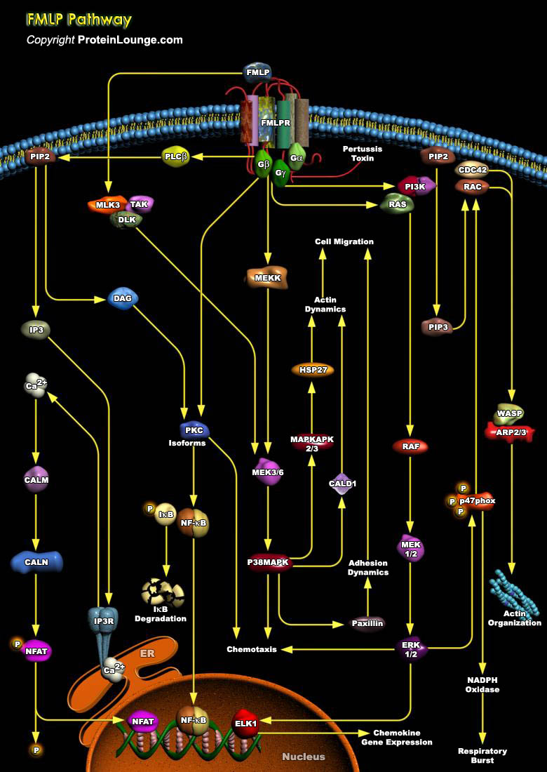
Neutrophils play an important role in the host defense by invading microbial pathogens. Upon infection neutrophils become activated through interaction with chemo attractants and cytokines. These ligands bind to a variety of cell surface receptors, including heterotrimeric GPCR (G-Protein Coupled Receptors) for fMLP (N-formyl-Met-Leu-Phe) and PAF (Platelet Activating Factor), and tyrosine kinase-associated receptors for GMCSF (Granulocyte-Macrophage Colony Stimulating Factor). Receptor activation triggers intracellular signal transduction pathways, resulting in the correct biological response, for instance, migration, phagocytosis, antibody-dependent cell mediated cytotoxicity, degranulation, superoxide production, transcriptional activation, and actin[..]
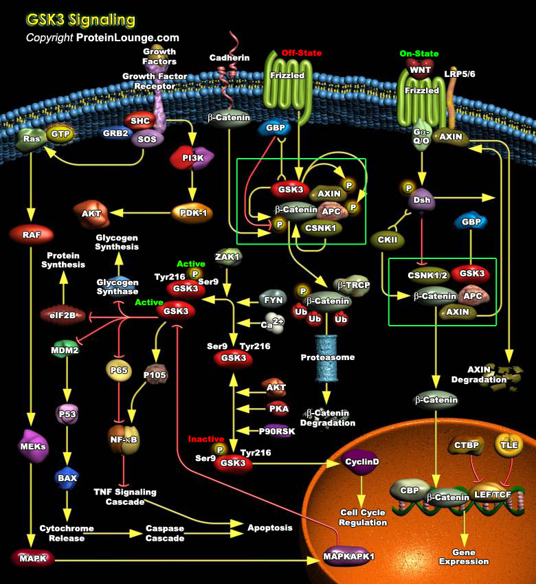
GSK3 (Glycogen Synthase Kinase-3) is a ubiquitously expressed, highly conserved serine/threonine protein kinase found in all eukaryotes. Identified originally as a regulator of glycogen metabolism, GSK3 acts as a downstream regulatory switch for numerous signaling pathways, including cellular responses to WNT, Growth Factors, Insulin, RTK (Receptor Tyrosine Kinases), Hedgehog pathways, and GPCR (G-Protein-Coupled Receptors) and is involved in a wide range of signal transduction cascades involving cellular processes, ranging from glycogen metabolism, cell development, gene transcription, protein translation to cytoskeletal organization, cell cycle regulation, proliferation and apoptosis. Unlike most protein kinases involved in signaling, GSK3 is active in unstimulated,[..]
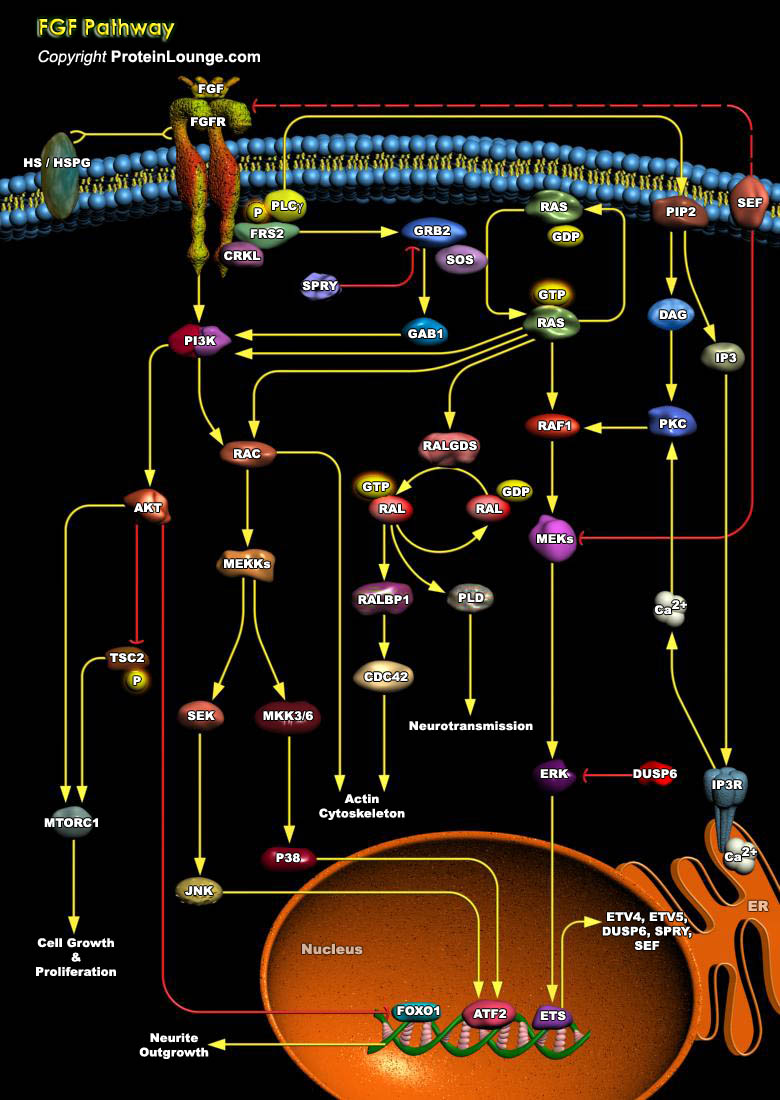
Angiogenesis, the growth of new blood vessels, plays a key role in many physiological and pathological processes, such as ovulation, embryogenesis, wound repair, inflammation, malignant tumor growth, retinopathies, rheumatoid arthritis, and angiogenesis-dependent diseases. One of the best-characterized modulators of angiogenesis is the heparin-binding FGF (Fibroblast Growth Factor) (Ref. 1).FGFs are a large family of multifunctional peptide growth factors of which there are at least 28 distinct members. The members of this peptide growth factor family have been identified in a variety of organisms and play pivotal roles in many cellular processes including mitogenesis, differentiation, migration, and cell survival During embryonic development, FGFs play a critical role[..]
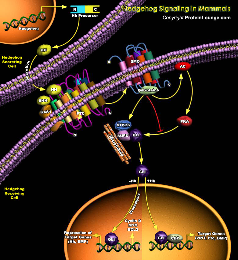
Controlled cell proliferation is a predominant theme in normal embryonic and post-embryonic development, and, in many instances, cell-type specification and cell proliferation are intimately coupled. Several secreted intercellular signaling proteins that behave as morphogens during pattern formation are also implicated in the regulation of the cell cycle. Hedgehogs (Hhs) are one such class of morphogens that regulate an enormous variety of developmental events in the fly and vertebrate embryo and plays a central role in several cancers.The vertebrate Hh family is represented by at least three members: Dhh (Desert Hh), Ihh (Indian Hh) and Shh (Sonic Hh), two Patched homologs, Ptc1 (Patched-1) and Ptc2 (Patched-2); and three homologs of Ci (Cubitus interruptus, a 155 kDa[..]