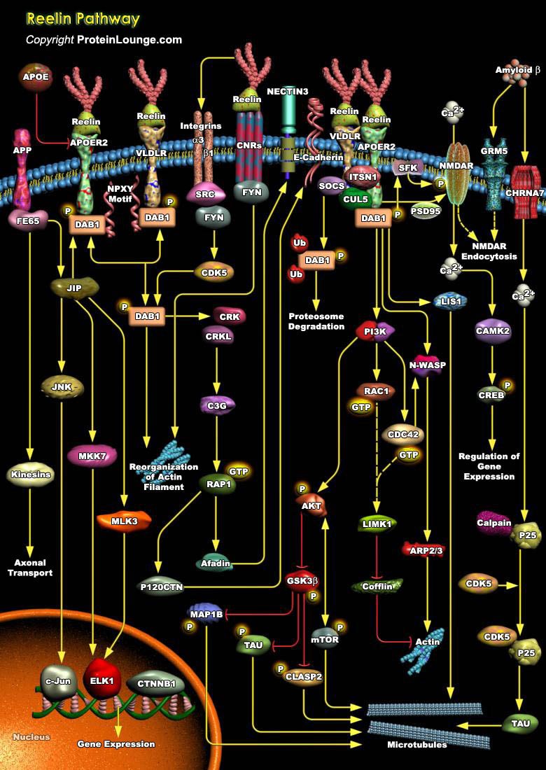
Reelin is a large extracellular glycoprotein involved in the development of architectonic patterns, particularly in the cerebral cortex and hippocampus, where primarily Cajal-Retzius cells synthesize it The Reelin signaling pathway controls neuronal migration and positioning in central nervous system (CNS) development, as well as synaptic plasticity and memory (Ref.1). Reelin is localized to chromosome 7 in human with serine protease activity containing 3461 amino acids. It contains a signal peptide followed by an N-terminal sequence and a hinge region upstream from eight Reelin repeats of 350–390 amino acids. Each Reelin consists of a signal peptide (S), an F-spondin-like domain (SL), eight consecutive Reelin repeats (R) each harboring an epidermal growth[..]
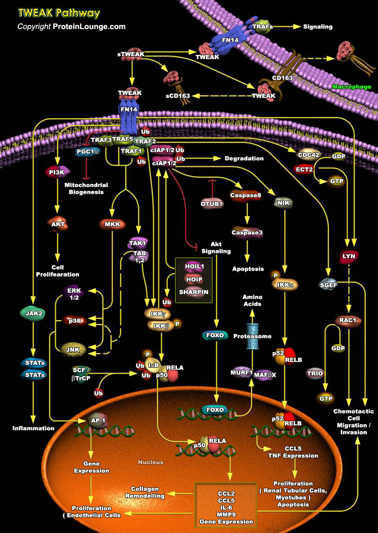
Tumor necrosis factor (TNF)-like weak inducer of apoptosis (TWEAK) is a member of the tumor necrosis factor (TNF) superfamily (TNFSF) that is induced in a variety of cell types in situations of tissue injury. It is expressed in various tissues including the intestine, pancreas, lung, brain, skeletal muscle, heart and vasculature. The 249 amino acid-comprising membrane-bound form of TWEAK consists extracellularly of the characteristic C-terminally located TNF homology domain (THD), which is separated from the transmembrane domain by a stalk region of about 55 amino acids. It can be cleaved by furin endoprotease into a soluble TWEAK (sTWEAK) (185 aa, 18 kDa) which then binds to the Fibroblast growth factor-inducible 14 (Fn14) receptor on the cell, the cognate receptor of[..]
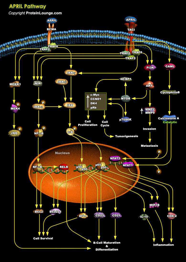
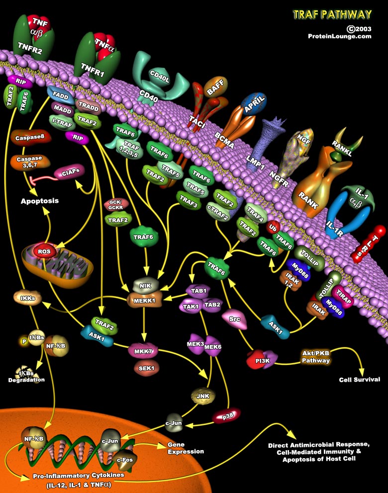
APRIL (a proliferation-inducing ligand, also known as TRDL-1, TALL-2, and TNFSF13), is a member of the TNF (tumor necrosis factor) superfamily, with homologous structure and function to several other cytokines in this family. It is a cytokine which is over-expressed by transformed cells and could stimulate cellular proliferation (Ref.1) It has two receptors i.e BCMA and TACI (Tumor necrosis factor receptor superfamily members 17 and 13B) that play important roles in the B-cell and T-cell arms of the immune system (Ref.2 & 3). One of the most important APRIL-induced mechanism is an activation of NF-kB (Nuclear factors of kappa light polypeptide in B-cells) signaling cascades. Activation and nuclear translocation of NF-kB proteins can occur by one of two pathways:[..]
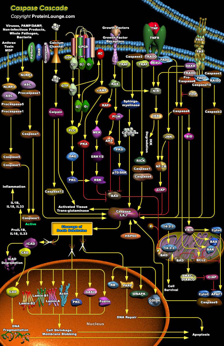
Caspases are a family of evolutionary conserved cysteine dependent aspartate-specific proteases that play crucial roles in maintaining organismal homoeostasis throughout life.Mammalian caspases are broadly classified as being inflammatory/pyroptotic (human caspase-1, 4, 5 and 12, murine caspase-1, 11 and 12), or as initiators (human and murine caspase-2, 8, 9 and human caspase-10) and executioners (human and murine caspase-3, 6 and 7) of apoptotic cell death.The culmination of this cascade is the cleavage of a number of proteins in the cell, followed by cell disassembly, cell death, and, ultimately, the phagocytosis and removal of the cell debris. The Caspase cascade is activated by two distinct routes: one from cell surface and the other from mitochondria (Ref.1). The[..]
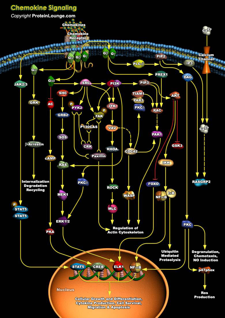
Chemokines are a group of small, secreted molecules that signal through G protein-coupled receptors to promote cell survival and proliferation and to provide directional guidance to migrating cells. Initially chemokines were divided into groups based on having chemotactic or homeostatic function, but several dual-function chemokines have since been described. To date, 44 chemokines and 23 chemokine receptors have been identified in the human genome. It is the expression of particular chemokines, receptors, and adhesion molecules that contribute to the selective migration and tissue specificity of leukocytes. Chemokines can be subdivided into four families based on the positioning of the N-terminal cysteine residues (Ref.1 & 2).Chemokines mediate their effects[..]
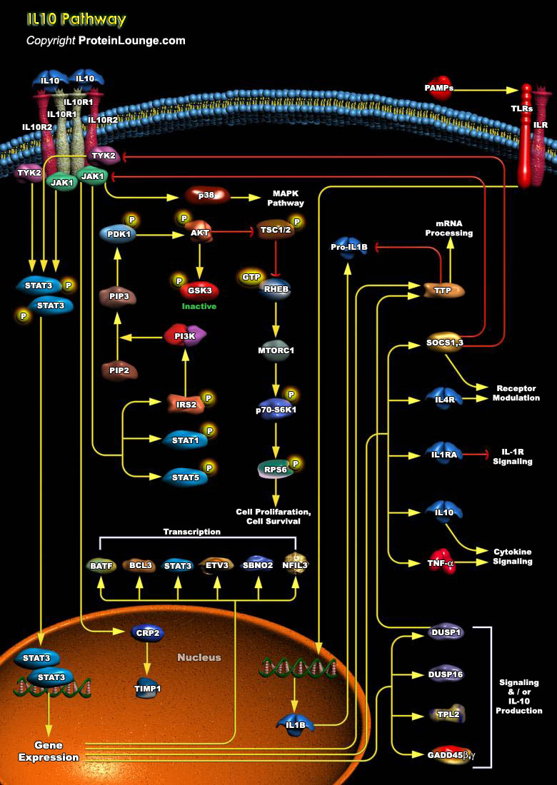
IL-10 (Interleukin-10) is a pleiotropic cytokine with important immunoregulatory functions whose actions influence activities of many of the cell-types in the immune system. It is a cytokine with potent anti-inflammatory properties, repressing the expression of inflammatory cytokines such as TNF-Alpha (Tumor Necrosis Factor-Alpha), IL-6 (Interleukin-6) and IL-1 (Interleukin-1) by activated macrophages (Ref.1). Functional IL-10R (IL-10 Receptor) complexes are tetramers consisting of two ligand-binding subunits (IL-10R-Alpha or IL-10R1) and two accessory signaling subunits (IL-10R-Beta or IL-10R2). Binding of IL-10 to the extracellular domain of IL-10R1 activates phosphorylation of the receptor-associated, JAK1 (Janus Kinase-1) and TYK2 (Tyrosine Kinase-2), which are[..]
To achieve strong adhesion to their neighbors and sustain stress and tension, epithelial cells develop many different specialized adhesive structures. Breakdown of these structures occurs during tumor progression with the development of a fibroblastic morphology characteristic of metastatic cells. Adhesion receptors of the Cadherin family have been implicated in these cellular processes, which play an important role in the development and maintenance of the differentiated epithelial phenotype during organogenesis and adult life. Cadherin-mediated adhesion requires the activity of the cytosolic proteins of the Rho subfamily members, Rho, Rac and CDC42 (Cell Division Cycle-42). They belong to the Ras Superfamily of small GTPases, whose function is regulated[..]
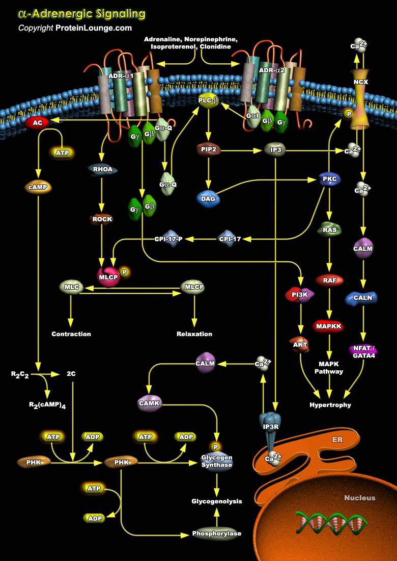
ADRs (Adrenergic Receptors) are expressed on virtually every cell type in the body and are the receptors for Adrenaline, Epinephrine and Norepinephrine within the Sympathetic Nervous System. They serve critical roles in maintaining homeostasis in normal physiologic settings as well as pathologic states. These receptors are also targets for therapeutically administered agonists and antagonists (Ref.1). ADRs are members of the super family of cell surface receptors that carry out signaling via GPCR (G-Protein Coupled Receptors) and are divided into nine distinct subtypes: ADR-Alpha1A, ADR-Alpha1B, ADR-Alpha1D, ADR-Alpha2A, ADR-Alpha2B, ADR-Alpha2C, ADR-Beta1, ADR-Beta2 and ADR-Beta3. ADR-Alpha2 is implicated in diverse physiological functions particularly of the[..]
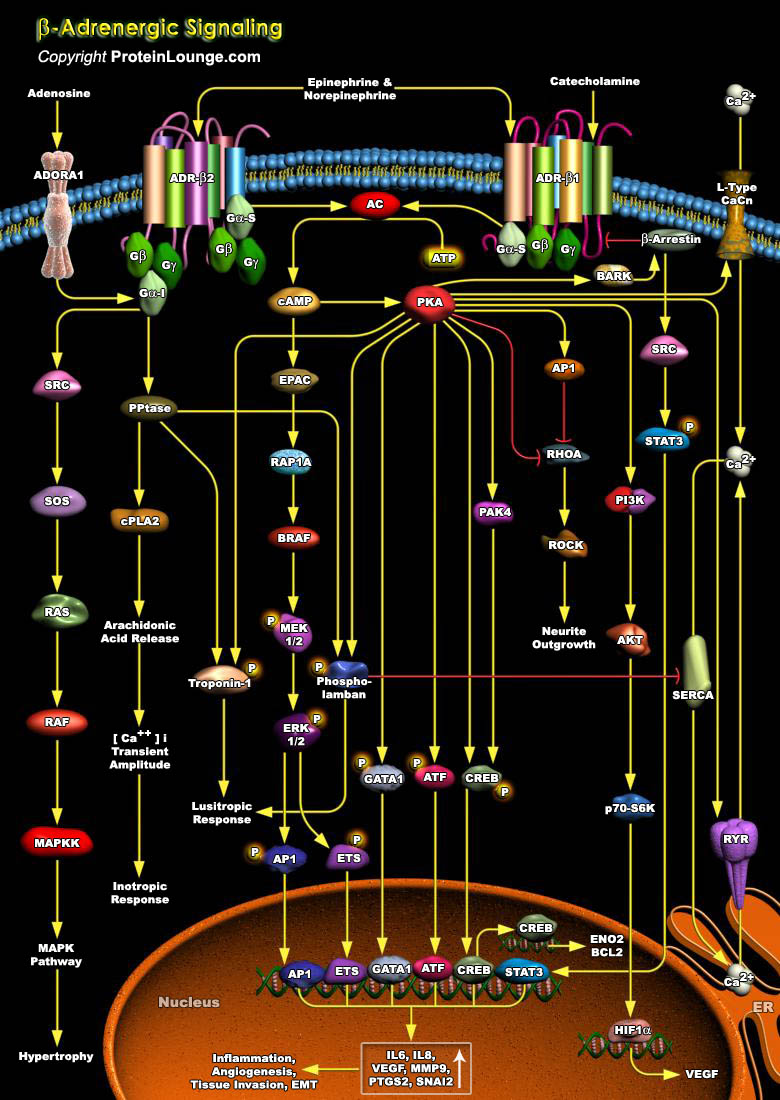
The rate and strength of beating of the heart is under the reciprocal control of the Adrenergic (sympathetic) and Cholinergic (parasympathetic) systems. Increased strength (inotropy) in cardiac beating in response to hormones like the blood-borne Epinephrine or to neurally delivered Norepinephrine is mediated by ADR-Beta (Beta-Adrenergic Receptors) , which are members of the superfamily of cell surface receptors that carry out signaling via coupling to G-proteins (Guanine nucleotide binding proteins)(Ref.1). ADR forms the interface between the endogenous Catecholamines, Adrenaline, Epinephrine and Norepinephrine and a wide array of target cells in the body to mediate the biological effects of the Sympathetic Nervous System. They serve critical roles in maintaining[..]
The structural and metabolic integrity of bone is maintained through the dynamic process of bone remodeling those results from the coordinate action of bone resorption and the formation of new bone by osteoblasts. Regulation of bone remodeling occurs through multiple mechanisms that ultimately converge on the interaction of osteoclasts or their precursors with osteoblasts and bone marrow stromal cells. Two key factors supplied by the stromal environment are CSF1 (Colony-Stimulating Factor-1) and the TNF family member, RANKL (Receptor Activator of Nuclear Factor-KappaB Ligand, also called TRANCE, ODF, OPGL). Signaling through RANK is essential for the differentiation and activation of osteoclasts, the cell principally responsible for bone resorption. RANK provokes[..]
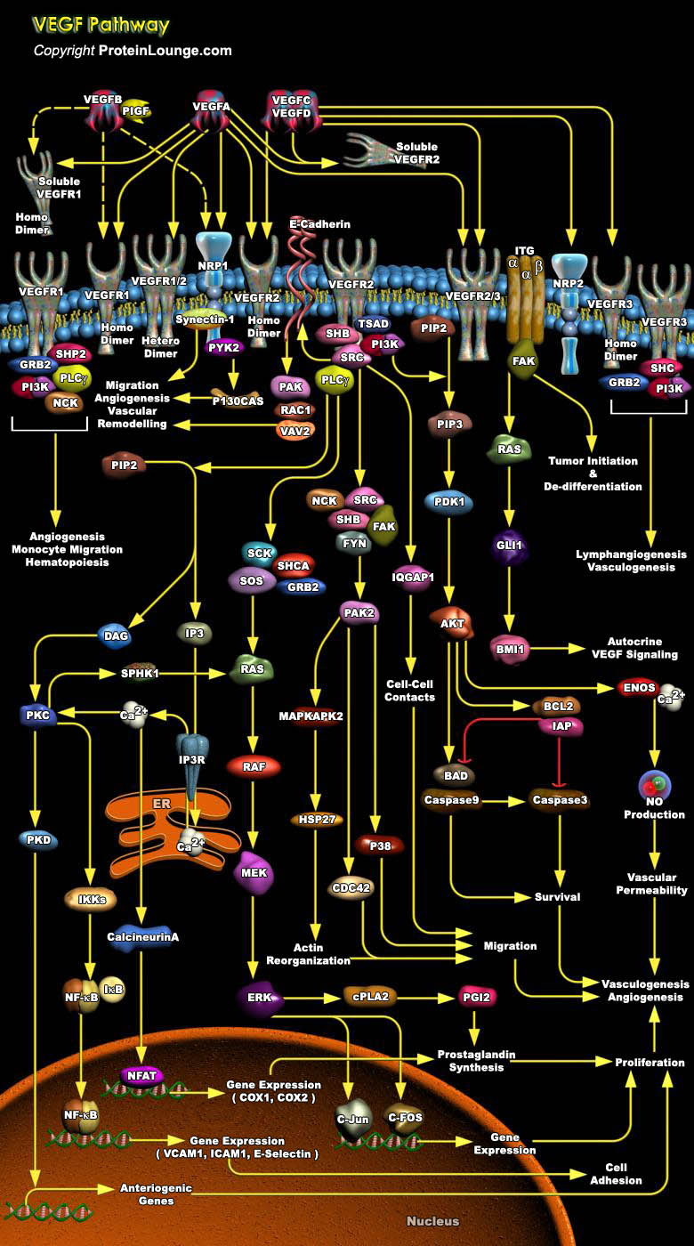
The vascular endothelial growth factor (VEGF) family of soluble protein growth factors is master regulators of in vasculogenesis and angiogenesis during blood vessel development. In mammals, the VEGF family consists of 5 members, VEGFA, B, C, and D and placenta growth factor (PIGF). VEGFs act through three structurally related VEGF receptor tyrosine kinases, denoted VEGFR1 (FLT1), VEGFR2 (FLK1), and VEGFR3 (FLT4). Binding of VEGF to its cognate VEGF receptor in cis or trans (e.g., by binding HS proteoglycans [HSPGs] on adjacent cells induces receptor homo- or heterodimerization (Ref.1 and 2). VEGFA binds VEGFR1 and VEGFR2. VEGFR2 is expressed mainly in endothelial cells, whereas VEGFR1 is expressed in endothelial cells as well as hematopoietic[..]
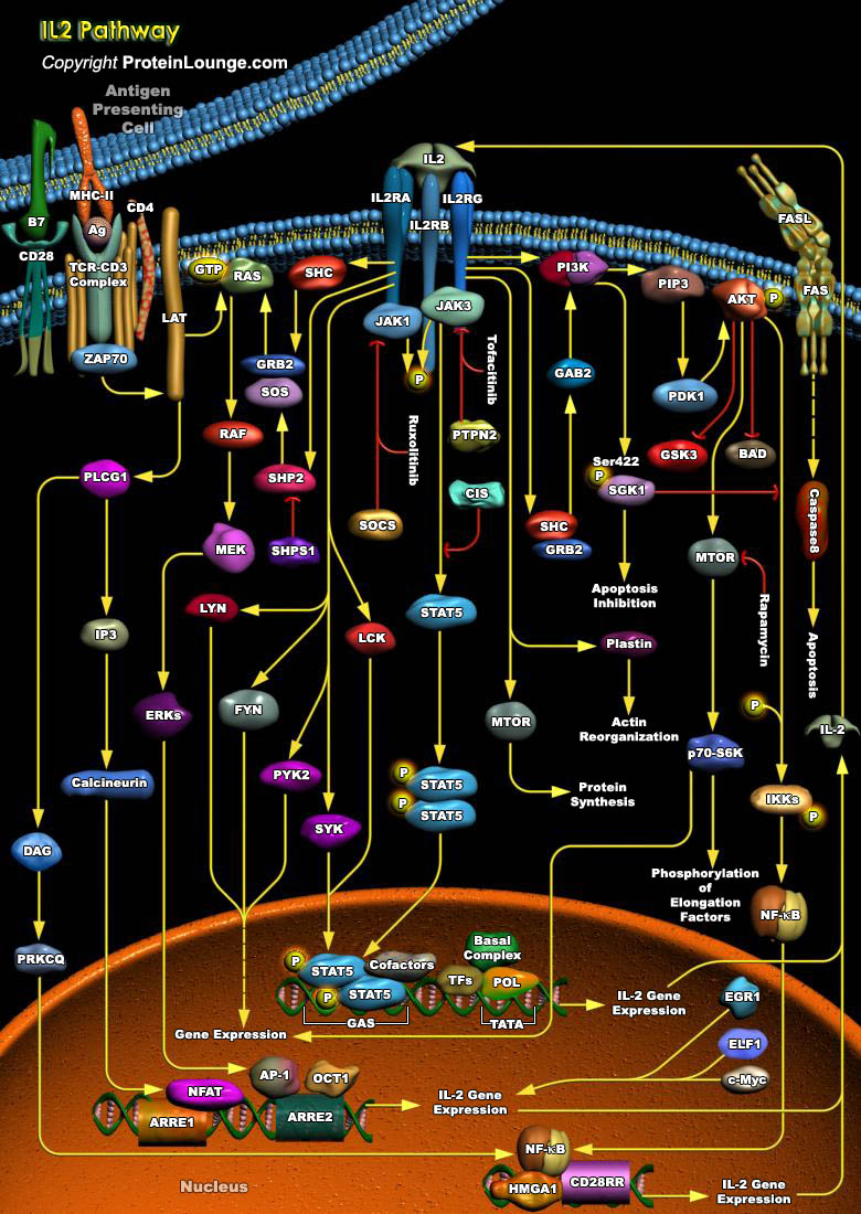
IL-2 (Interleukin-2) is a T-Cell-derived cytokine important in the regulation of growth and differentiation of T-Cells, B-Cells, natural killer cells, glioma cells, and cells of the monocyte lineage after specifically interacting with its receptors. Human IL-2 is a 133-amino acid polypeptide with a molecular mass of 15-18 kDa. IL-2 signaling is mediated by a multichain receptor complex consisting of an alpha (CD25), beta (CD122) and gamma (CD132) chain. The IL-2R (IL-2 Receptor) alpha subunit primarily increases the affinity of ligand binding and is not known to contain a signaling domain, whereas the beta and gamma subunits participate in both ligand binding and signal transduction. The IL-2R signaling system proceeds through at least three different pathways, which[..]