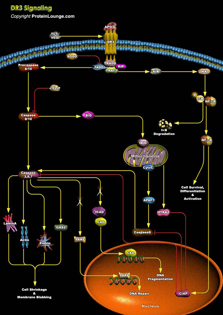
The TNFR (Tumor Necrosis Factor Receptor) superfamily comprises a growing family of type I membrane bound glycoproteins, which interact with the TNF family of soluble mediators and type II transmembrane proteins. At least 23 TNFR superfamily members and 17 known ligands have been identified in mammals. These receptors trigger pleiotropic responses, ranging from apoptosis and differentiation to proliferation, and have been implicated in immune regulation, host defense and lymphoid organ development. Members of the TNFR family are characterized by the presence of varying numbers (two to six) of cysteine-rich repeats in their cytoplasmic domains. Among these molecules, a novel subgroup has been defined, termed DR (Death Receptors), as one of their most prominent functions[..]
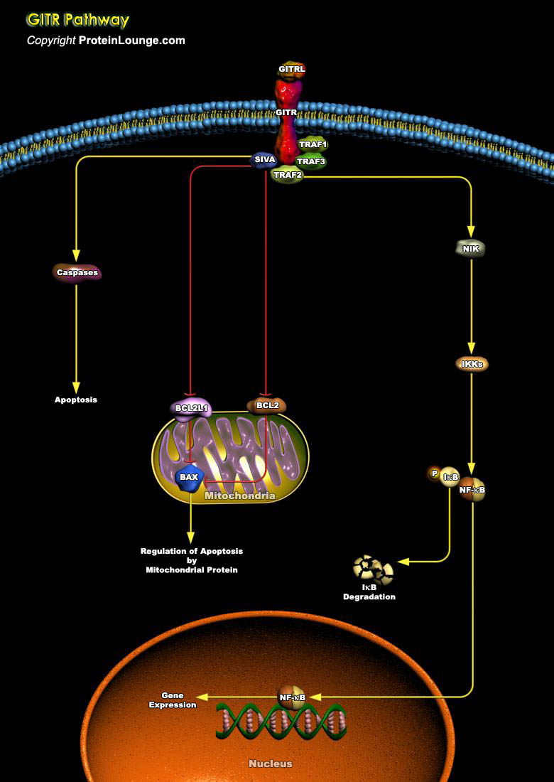
GITR (Glucocorticoid-Induced TNFR Family-Related) also termed AITR (Activation-Inducible TNFR Family Receptor) is a member of the TNFRSF18 (TNF Receptor Superfamily 18). It is a 228-amino acids type I transmembrane protein that is suggested to be a close relative of 4-1BB and CD27. Inducible during T-Cell activation, the molecule has a 19 amino acid residue signal sequence, a 134 amino acid residue extracellular region, a 23 amino acid residue transmembrane segment and a 52 amino acid residue cytoplasmic domain. It has three cysteine-rich motifs in its extracellular region. Its ligand is GITRL (AITRL). GITR expression is upregulated on T-Cells. A high level of GITR is constitutively expressed on CD4+ CD25+ regulatory T-Cells. CD4+ GITR+ T-Cells are equivalent to CD4+[..]
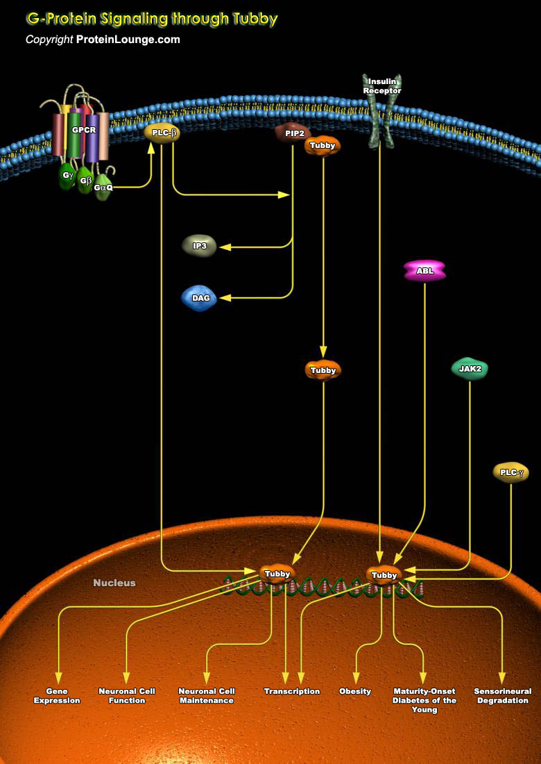
The Tubby protein is the founding member of a multigene protein family that plays an important role in maintenance and function of neuronal cells during development and post-differentiation. Currently, four Tubby gene family members (TUB, TULP1, TULP2 and TULP3) have been identified, which are conserved among different species of mammals (Ref.1). Besides, Tubby-like proteins are also found in other multicellular organisms including plants. These proteins feature a characteristic "Tubby domain" of approximately 260 amino acids at the C-terminus that forms a unique helix-filled barrel structure; this C-terminal domain binds avidly to double-stranded DNA. Most Tubby proteins include NH2-terminal regions that, in general, are not closely related to one another.[..]
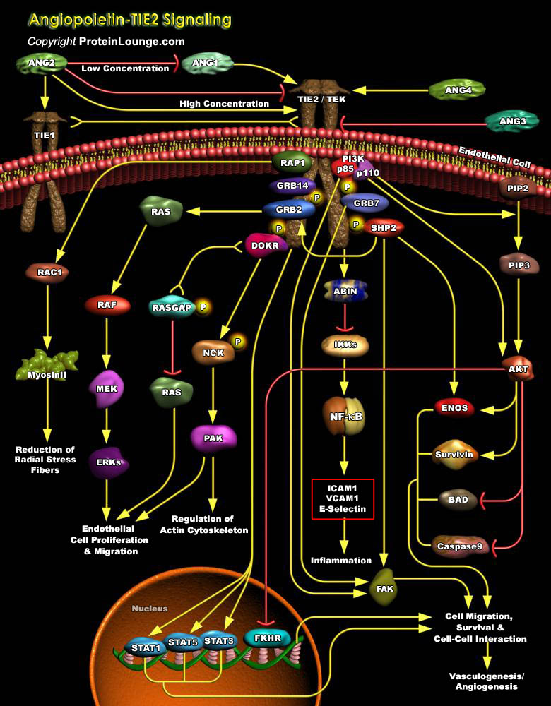
Development of a functional cardiovascular system is dependent on the regulated proliferation, migration, and differentiation of endothelial cells in two discrete processes known as vasculogenesis and angiogenesis. Angiogenesis is the formation of new capillaries from pre-existing vessels, whereas vasculogenesis is de-novo capillary formation from EPCs (Endothelial Precursor Cells). New capillaries arise from preexisting larger vessels to give rise to a more complex vascular network with a hierarchy of both large and small vessels (Ref.1). These sequential vascular developments are tightly regulated by a range of pro- and antiangiogenic factors, including VEGF (Vascular Endothelial Growth Factor ), bFGF(basic Fibroblast Growth Factor ), Thrombospondin, Angiopoietins,[..]
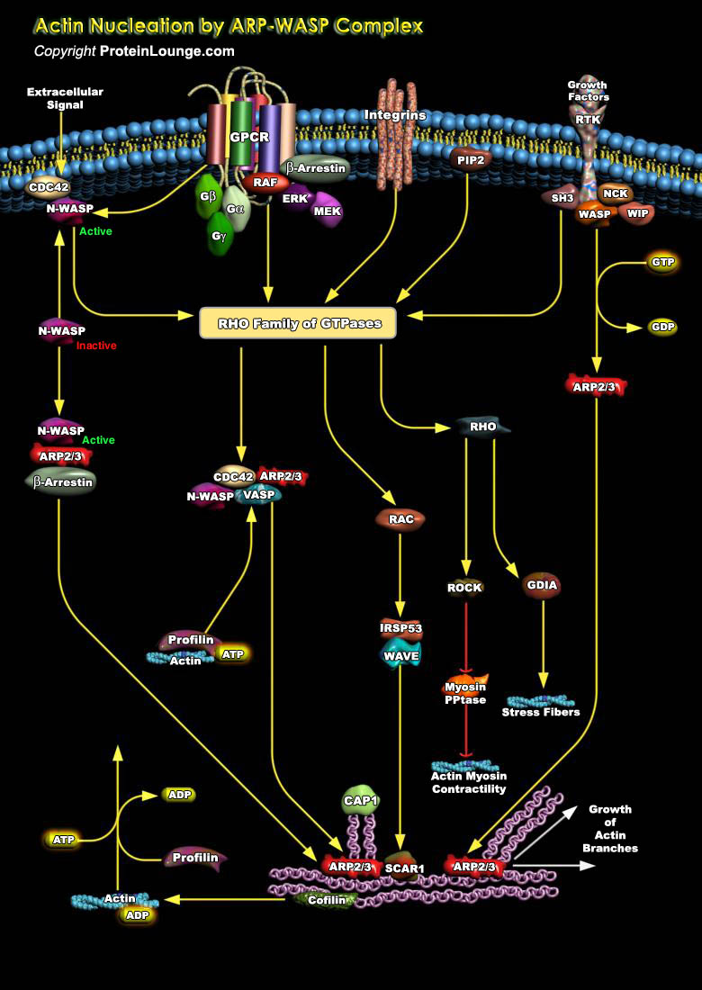
Actin Nucleation By ARP-WASP Complex For many cell types, the ability to move across a solid surface is fundamental to their biological function. Certain aspects of cell locomotion, such as the protrusion of the plasma membrane in lamellipodia and filopodia, are driven by the polymerization of actin cytoskeleton. The actin cytoskeleton is a dynamic filament network that is essential for cell movement during embryo development, polarization, morphogenesis, cell division, and immune system function and in the metastasis of cancer cells. To engage in these complex behaviors, cells must direct actin assembly with a high degree of spatial and temporal resolution in response to extracellular signals (Ref.1). To coordinate these behaviors, tight spatial and temporal control[..]
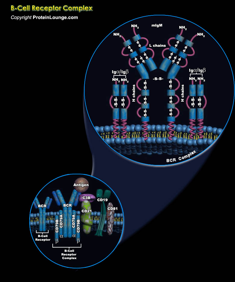
Lymphocytes are one of the five kinds of white blood cells or leukocytes, circulating in the blood. Although mature lymphocytes all look pretty much alike, they are extraordinarily diverse in their functions. The most abundant lymphocytes are: B-Lymphocytes (often simply called B-Cells) and T-Lymphocytes (likewise called T-Cells) [Ref.1]. B-Cells are not only produced in the bone marrow but also mature there. Each B-Cell is specific for a particular antigen. The specificity of binding resides in the BCR (B-Cell receptor) for antigen [Ref.2]. Mature B cells express two BCR isotypes, IgM and IgD. The BCR is composed of mIg (membrane immunoglobulin); a structure of four (in the case of IgD) or five (IgM) immunoglobulin domains in the heavy chain linked by a hinge, and a[..]
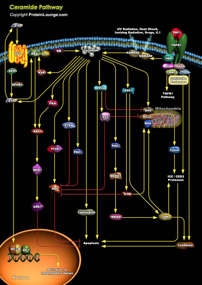
The SM (Sphingomyelin) pathway is an evolutionarily conserved stress response system linking diverse environmental stresses (Ultraviolet, Heat Shock, Oxidative Stress, and Ionizing Radiation) to cellular effector pathways. Ceramide is the second messenger in this system and can be generated either by hydrolysis of SM through SM-specific PLC (Phospholipase-C) termed SMases (Sphingomyelinases) or by de novo synthesis through the enzyme Ceramide Synthase . There are two classes of SMase, acidic (A-Smase) and neutral (N-Smase). Stress stimuli such as, TNF-Alpha (Tumor Necrosis Factor-Alpha), lipopolysaccharide, and some chemotherapy drugs such as doxorubicin also mediate apoptosis by generation of the lipid second messenger, Ceramide. A specific interaction between the FAN[..]

The ability of multicellular organisms to maintain cellular homeostasis is critically dependent on a balance between cell survival and cell death (apoptosis). The responsiveness of individual cells to death signals varies greatly depending on the presence of continuous survival cues from the extracellular environment. The perturbation of normal cell survival mechanisms, leading to an increase in cell survival or cell death plays an important role in the development of a number of disease states, including cancer. BAD (BCL2 Associated Death Promoter) is a pro-apoptotic critical regulatory component of the intrinsic cell death machinery that exerts its death-promoting effect upon heterodimerization with the antiapoptotic proteins of the BCL2 family defined by conserved[..]
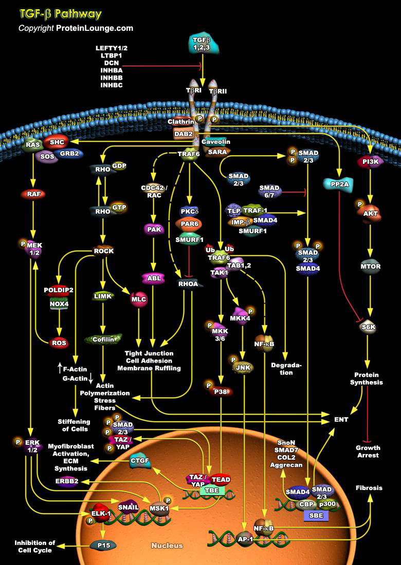
TGFB (Transforming growth factor-beta) is a multifunctional cytokine that regulates a wide variety of cellular functions, including cell growth, cellular differentiation, apoptosis, and wound healing. TGF-b signals are transmitted through two transmembrane serine/threonine kinase receptors TGFBR1 and TGFBR2 [Ref.1]. Initiation of the TGFB signaling cascade occurs upon ligand binding to TGFB receptor TGFBR2 and subsequent TGFBR1– TGFBR2 heterotetrameric complex formation. TGFBR2 is a constitutively active receptor kinase and phosphorylates Ser/Thr residues in the cytoplasmic GS domain of TGFBR1, which turns on the kinase activity of TGFBR1. Upon Activation, TGFBR1 transmits its signal to the various intracellular SMAD-dependent and SMAD independent signaling[..]
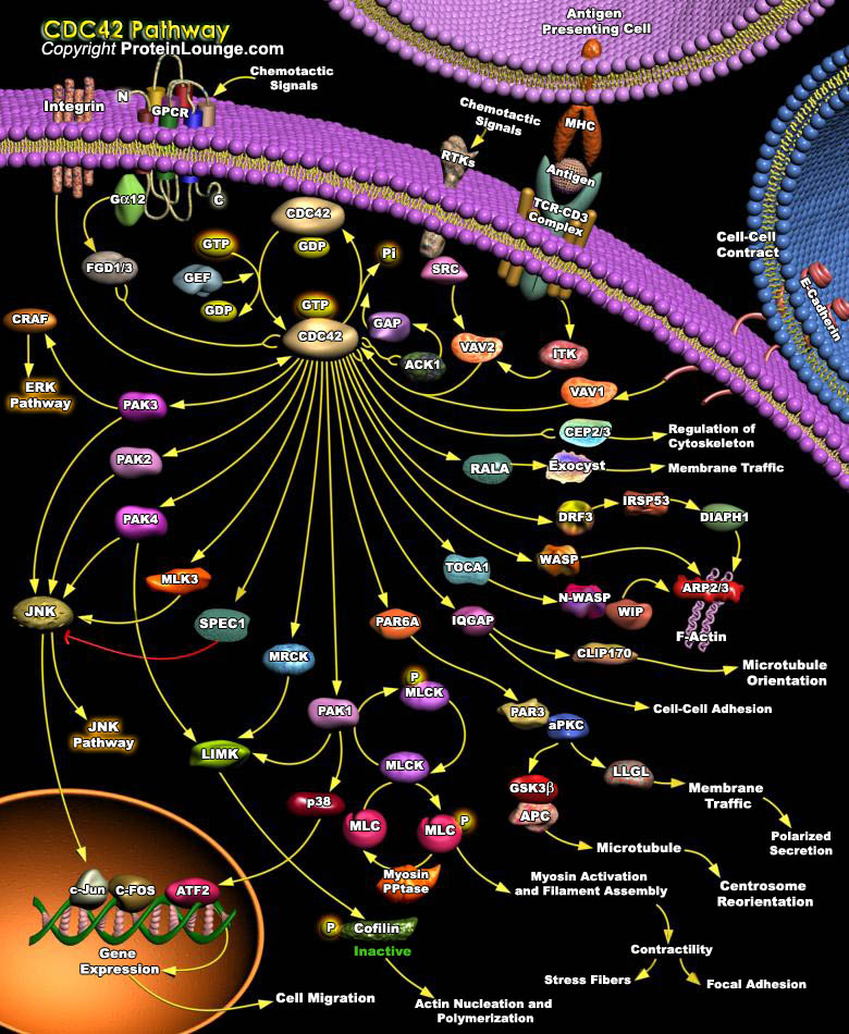
Dynamic regulation of the actin cytoskeleton underpins a multitude of cellular processes, from cell movement and polarization1,2 to cell division.3 The actin cytoskeleton and the proteins involved in its regulation are also fundamentally linked to endocytosis and membrane trafficking.4,5 Members of the Rho family of small GTPases have emerged as important overseers of the actin cytoskeleton and a number of Rho family members and their downstream effector proteins have been linked to specific actin pathways. Among the Rho family actin regulators is Cdc42 is a small GTPase responsible for a large number of eukaryotic cell signaling pathways(Ref.1).CDC42 acts downstream of cell surface receptors to regulate the formation of different F-actin-containing structures. It[..]
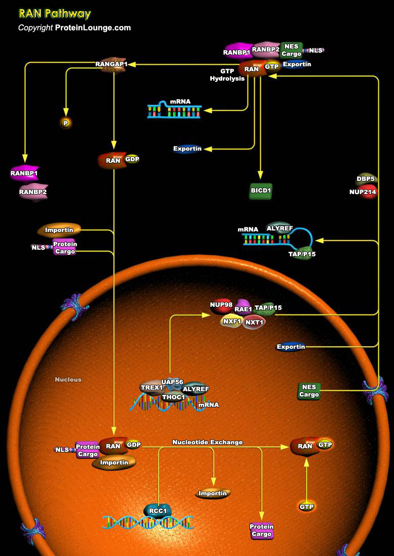
Ran is a member of the Ras family of small GTPases. The Ran subgroup is represented by its lone member, Ran, that is distinguished from Ras GTPases by its lipid modification and atypical subcellular localization. Unlike most other Ras-related proteins, Ran is not modified to bind to cell membranes. Instead, Ran protein is localized throughout the cell, where it is concentrated primarily in the nucleus (Ref.1). Ran is regulated by a cytosolic RanGAP1 (Ran GTPase–Activating Protein-1) and by a RanGEF (Chromatin-Bound Guanine Nucleotide Exchange Factor). The distribution of RanGTP provides important spatial information that directs cellular activities during different parts of the cell cycle. During interphase, the localization of RanGEF and RanGAP1 predicts that[..]
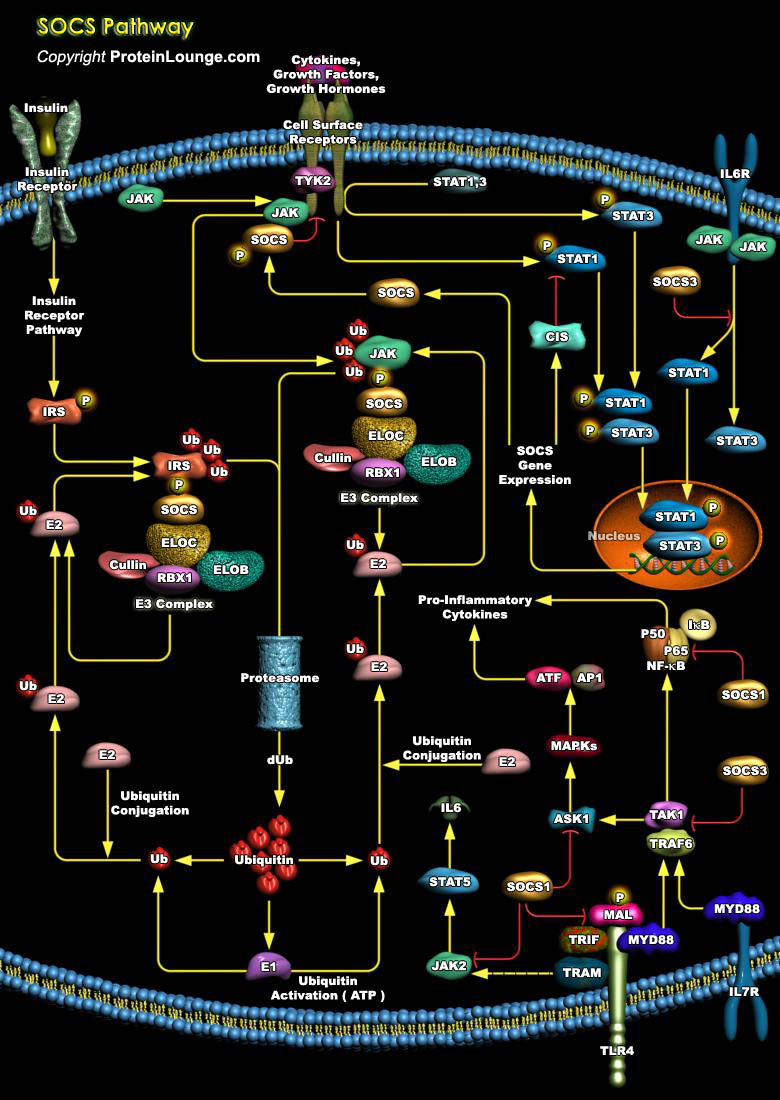
Our immune system is largely controlled by the action of pleiotropic cytokines and growth factors, small secreted proteins, which bind to receptors on the surface of immune cells to initiate an appropriate physiological response. The cellular response to cytokines and growth factors is predominantly executed by intracellular proteins known as the Janus kinases (JAKs) and the signal transducers and activators of transcriptions (STATs). These kinases activated upon ligand binding causes the phosphorylation of tyrosine residues within the receptor intracellular region and phosphorylation of other signalling proteins recruited to the receptor complex like signal transducers and activators of transcriptions (STATs). Once phosphorylated, STATs translocate to the[..]

